OVERVIEW

What Is the skeleton System?

skeleton Muscles

Theskeleton muscles are the most common type of muscle tissue in the human body, accounting for approximately 30–40% of total body mass. They are one of the three types of muscles, the other two being cardiac muscle and smooth muscle. As part of the musculoskeleton system, skeleton muscles are attached to bones by strong connective tissues called tendons. They are made up of long, cylindrical muscle fibers grouped into bundles known as fascicles, with each fiber containing numerous myofibrils composed of the proteins actin and myosin. These muscles have a characteristic striped (striated) appearance under the microscope due to the orderly arrangement of their contractile proteins. skeleton muscles enable voluntary movement, help maintain body posture, stabilize and protect joints, and generate heat to help regulate body temperature. Their complex structure and ability to contract under conscious control make them essential for nearly every physical activity, from simple everyday movements to intense exercise.
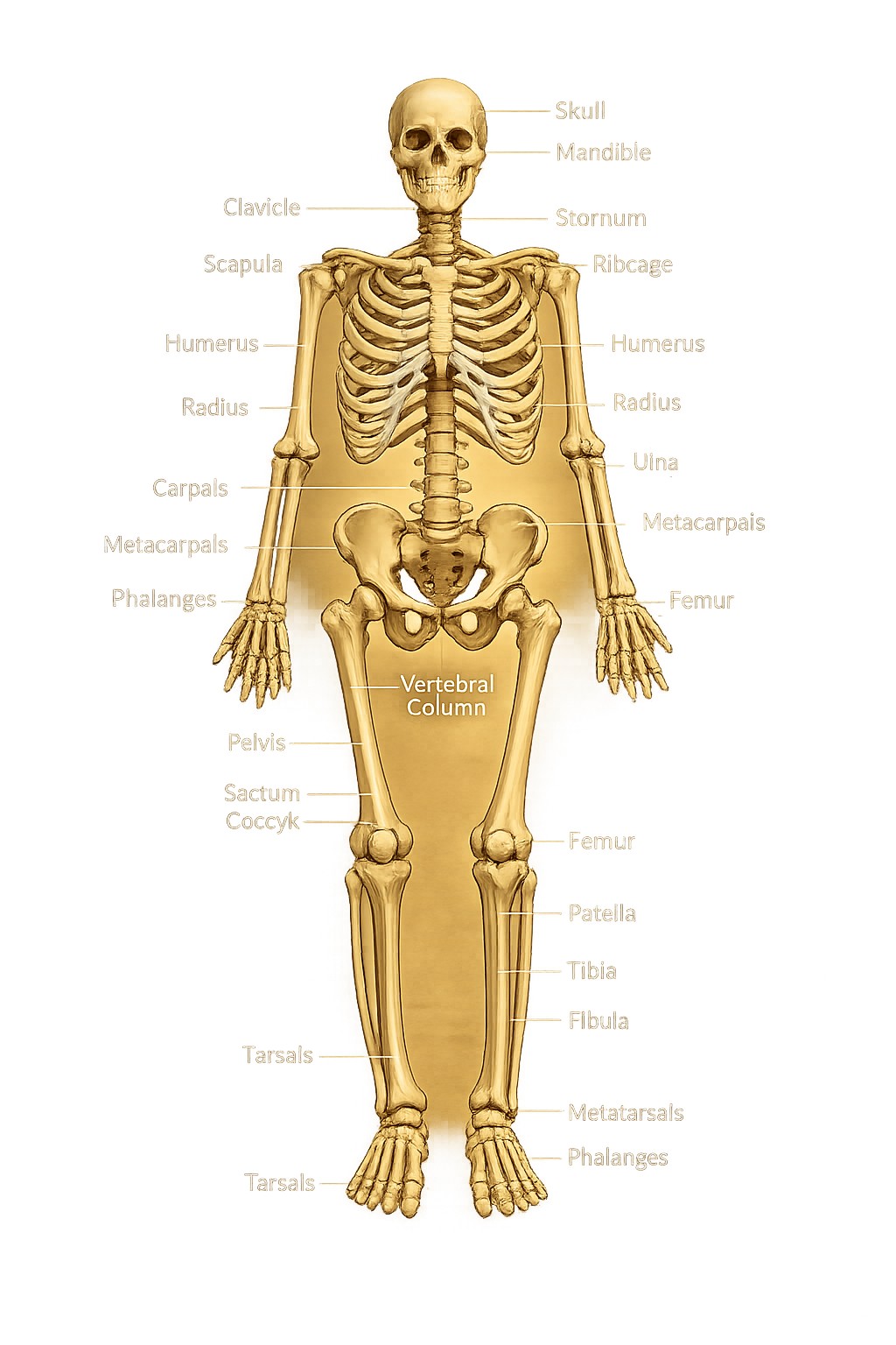
Interactive 3D Human skeleton System

Rotate, zoom and inspect the human skeleton System to explore its structure from every angle.

skeleton System Labelled Illustrations

skeleton System

Human skeleton System

Divisions of the Reproductive System

Axial Skeleton

The axial skeleton forms the central axis of the body and consists of the skull, vertebral column, ribs, and sternum. It provides support, maintains posture, and protects the body's vital organs.

Appendicular Skeleton

The appendicular skeleton consists of the bones of the upper limbs, lower limbs, shoulder girdle, and pelvic girdle. It enables body movement, supports locomotion, and connects the limbs to the axial skeleton.

Bone Classification

Bones are classified according to their shape rather than their size. Each type of bone has a unique structure that allows it to perform specific functions such as movement, protection, support, or reducing friction.

🦴 Long Bones

Long bones are longer than they are wide. They act as strong levers that support body weight and help produce movement. Most long bones are found in the arms and legs.

Examples: Femur, Humerus, Tibia, Fibula, Radius, Ulna.

🦴 Short Bones

Short bones are nearly equal in length, width, and thickness. They provide stability, support, and limited movement while absorbing shock.

Examples: Carpals (wrist bones) and Tarsals (ankle bones).

🦴 Flat Bones

Flat bones are thin and often slightly curved. Their broad surfaces protect delicate organs and provide large areas for muscle attachment.

Examples: Skull bones, Sternum, Ribs, Scapula.

🦴 Irregular Bones

Irregular bones have complex shapes that do not fit into the other categories. They provide protection, support, and multiple muscle attachment sites.

Examples: Vertebrae, Sacrum, Coccyx, Hip Bones.

🦴 Sesamoid Bones

Sesamoid bones develop inside tendons. They protect tendons from wear, reduce friction, and improve the efficiency of muscle movement.

Example: Patella (kneecap).

Structure of a Long Bone

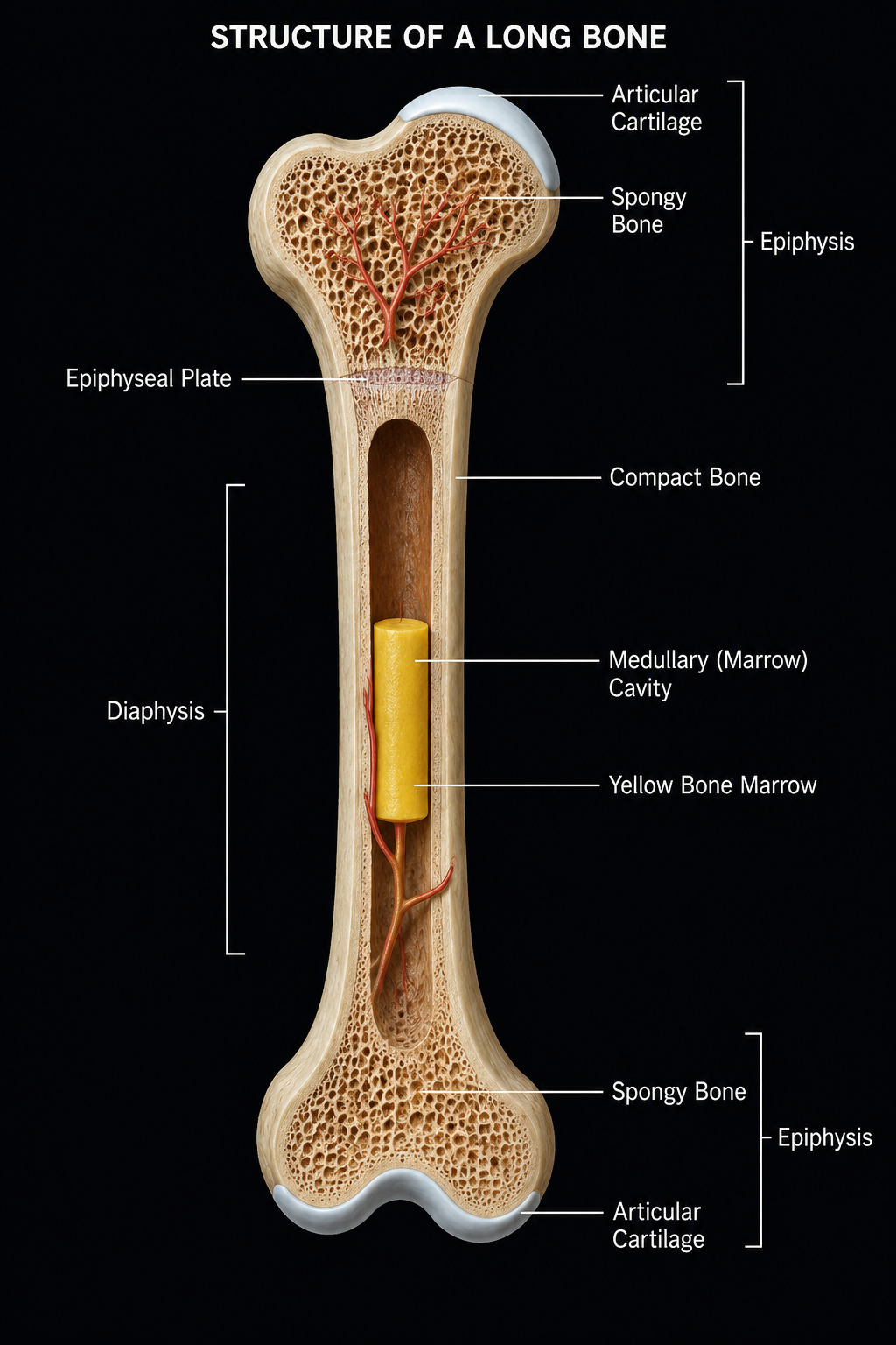
A typical long bone consists of several specialized regions that work together to provide strength, flexibility, protection, and blood cell production.

Structure of a Long Bone

🦴 Epiphysis

The enlarged ends of a long bone are called the epiphyses. They are mainly composed of spongy bone and are covered by articular cartilage, helping form smooth joints and absorb shock.

🦴 Diaphysis

The diaphysis is the long cylindrical shaft of the bone. It is made mostly of compact bone, providing strength and support while protecting the marrow cavity.

🦴 Compact Bone

Compact bone is the dense, hard outer layer of bone. It provides durability, supports body weight, and protects the internal structures of the bone.

🦴 Spongy Bone

Spongy bone, also known as cancellous bone, is a porous tissue found mainly inside the epiphyses. It reduces bone weight while maintaining strength and contains red bone marrow.

🦴 Bone Marrow

The marrow cavity contains bone marrow. Red bone marrow produces blood cells, while yellow bone marrow stores fat and serves as an energy reserve.

🦴 Periosteum

The periosteum is a tough, fibrous membrane that covers the outer surface of the bone, except at the ends where articular cartilage is present. It contains blood vessels and nerves that nourish the bone, protect it from injury, and help repair fractures. The periosteum also provides attachment points for tendons and ligaments.

Bone Formation (Ossification)

Ossification is the process by which bones are formed and developed. During fetal development and childhood, cartilage is gradually replaced by bone tissue until a mature bone is formed.

🌱

Mesenchyme

Bone development begins with mesenchymal cells, which are unspecialized embryonic cells capable of developing into various connective tissues.

⬇
🦴

Cartilage Model

Most long bones first develop as a cartilage model that provides the initial shape and framework of the future bone.

⬇
βš™οΈ

Primary Ossification

The primary ossification center develops in the diaphysis (shaft), where cartilage is gradually replaced by compact bone.

⬇
✨

Secondary Ossification

Secondary ossification centers appear in the epiphyses (ends) of the bone, allowing both ends to develop properly while growth continues.

⬇
πŸ›οΈ

Mature Bone

After complete ossification, the bone reaches its mature structure with compact bone, spongy bone, marrow cavity, and fully developed epiphyses.

Functions of the skeleton System

The skeleton system is much more than a framework of bones. It supports the body, protects vital organs, enables movement, produces blood cells, stores minerals, and performs several other essential functions that help maintain overall health.

🦴 Support

The skeleton forms the body's framework, giving it shape and providing support for muscles, tissues, and internal organs.

πŸ›‘οΈ Protection

Bones protect delicate organs. The skull protects the brain, the vertebral column protects the spinal cord, and the rib cage protects the heart and lungs.

πŸƒ Movement

Bones work together with muscles and joints to produce body movements such as walking, running, lifting, and jumping.

🩸 Blood Cell Formation

Red bone marrow inside certain bones produces red blood cells, white blood cells, and platelets through a process called hematopoiesis.

πŸ§ͺ Mineral Storage

Bones store important minerals such as calcium and phosphorus and release them into the bloodstream when the body requires them.

βš–οΈ Fat Storage

Yellow bone marrow stores fat, which serves as an energy reserve that the body can use when needed.

πŸ’ͺ Muscle Attachment

Bones provide attachment sites for muscles, tendons, and ligaments, allowing muscles to pull on bones and produce movement.

🧍 Maintains Posture

The skeleton system provides a stable framework that helps the body maintain balance, posture, and an upright position.

Bone Marrow

Bone marrow is a soft, spongy tissue found inside many bones. It plays a vital role in producing blood cells and storing energy. There are two main types of bone marrow, each with a unique function in maintaining the body's health.

🩸 Red Bone Marrow

Red bone marrow is responsible for hematopoiesis, the process of producing blood cells. It continuously forms red blood cells, white blood cells, and platelets, ensuring the body receives oxygen, fights infections, and repairs damaged blood vessels. Red marrow is mainly found in flat bones such as the sternum, ribs, pelvis, skull, and in the spongy ends of long bones.

🟑 Yellow Bone Marrow

Yellow bone marrow is composed mainly of fat cells and serves as the body's energy reserve. It is located primarily in the medullary cavity of long bones. During severe blood loss or certain medical conditions, yellow marrow can convert into red marrow to help increase blood cell production.

Classification of Joints

Joints are places where two or more bones meet. They hold the skeleton together while allowing different degrees of movement. Based on their structure and mobility, joints are classified into three main types.

🦴 Fibrous Joints

Fibrous joints are connected by dense connective tissue and allow little or no movement. They provide maximum strength and stability, making them ideal for protecting delicate organs.

Movement: Immovable (or nearly immovable)

Examples: Sutures of the skull and the joint between the tibia and fibula.

🦴 Cartilaginous Joints

Cartilaginous joints are connected by cartilage. They allow limited movement while providing flexibility and absorbing shock between bones.

Movement: Slightly movable

Examples: Intervertebral discs and the joint between the ribs and sternum.

🦴 Synovial Joints

Synovial joints are the most common and freely movable joints in the human body. They contain a synovial cavity filled with lubricating fluid that reduces friction and allows smooth movement.

Movement: Freely movable

Examples: Shoulder, elbow, wrist, hip, knee, ankle, and finger joints.

Types of Synovial Joints

Synovial joints are freely movable joints that allow a wide variety of movements. Depending on the shape of the articulating bones and the type of movement they permit, they are classified into six main types.

⚽ Ball & Socket

Allows movement in all directions, including rotation. It provides the greatest range of motion of any joint.

Examples: Shoulder and Hip.

πŸšͺ Hinge

Allows movement in one plane, similar to the opening and closing of a door.

Examples: Elbow, Knee, Fingers.

πŸŒ€ Pivot

Allows one bone to rotate around another, producing rotational movement.

Examples: Atlas-Axis joint and Radius-Ulna.

🐎 Saddle

Allows movement in two directions while providing greater flexibility than condyloid joints.

Example: Base of the Thumb.

🎯 Condyloid

Allows movement in two planes including flexion, extension, abduction, and adduction, but not rotation.

Examples: Wrist and Knuckles.

🧩 Gliding

Allows bones to slide over one another in various directions with limited range of movement.

Examples: Carpals, Tarsals, and Vertebral Facet Joints.

Hormonal Control of Bones

Several hormones help regulate bone growth, repair, and mineral balance throughout life.

🧬 Growth Hormone (GH)

Stimulates bone growth during childhood and adolescence by promoting the growth of cartilage and long bones.

🦴 Calcitonin

Lowers blood calcium levels by encouraging calcium to be deposited into bones, making them stronger.

βš–οΈ Parathyroid Hormone (PTH)

Raises blood calcium levels by stimulating the release of calcium from bones when the body needs it.

β˜€οΈ Vitamin D

Increases calcium absorption from the intestine and helps maintain healthy, strong bones.

Comparison: Compact Bone vs Spongy Bone

Although both compact and spongy bone are made of the same living tissue, they differ in structure, location, and function. Together they provide bones with both strength and reduced weight.

Feature Compact Bone Spongy Bone
Structure Dense and solid Porous and honeycomb-like
Location Outer layer of bones Inside bones, especially epiphyses
Strength Very strong and rigid Lightweight but supportive
Function Provides protection and support Absorbs shock and reduces bone weight
Bone Marrow Surrounds the marrow cavity Contains red bone marrow
Appearance Smooth and compact Network of tiny spaces (trabeculae)

Interesting Statistics

Here are some fascinating statistics that highlight the incredible strength and importance of the human skeleton system.

206

Bones in an Adult Skeleton

270

Bones in a Newborn Baby

80

Bones in the Axial Skeleton

126

Bones in the Appendicular Skeleton

99%

of the Body's Calcium is Stored in Bones and Teeth

2 Million

Red Blood Cells Produced Every Second by Bone Marrow

10 Years

Approximate Time to Completely Remodel Most Bones

5Γ—

Healthy Bone is Approximately Five Times Stronger than Steel (by Weight)

25%

The Femur Makes Up About One-Quarter of a Person's Height

3 mm

Length of the Smallest Bone (Stapes) in the Human Body

Did You Know?

🦴