Interactive Nerves present in our body Labelled Illustration
🟢 Dorsal Root (Posterior Root)
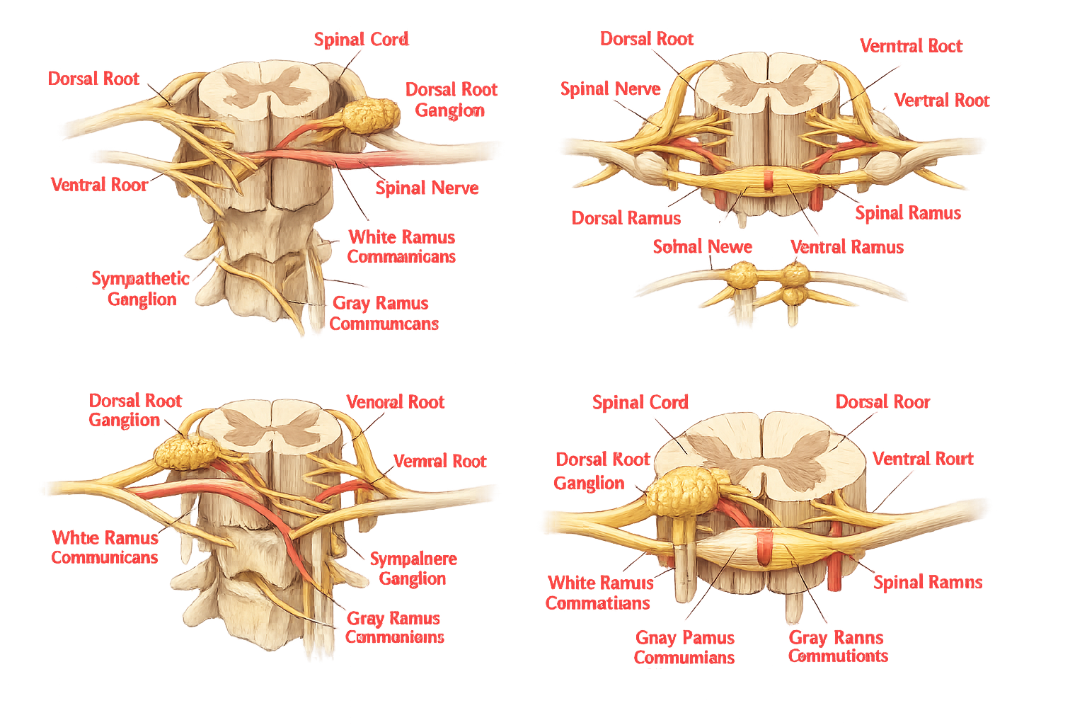
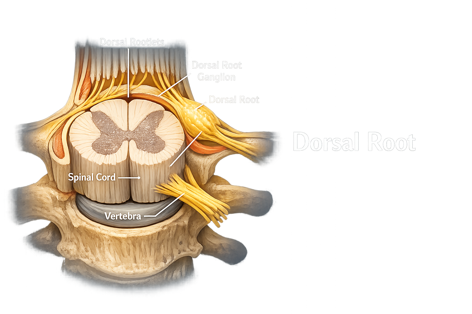
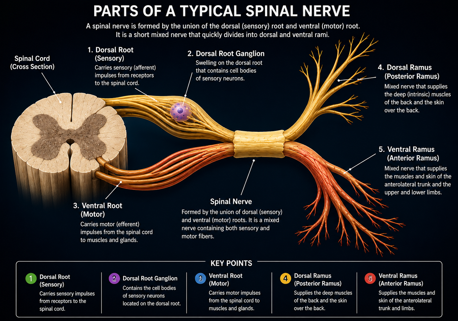
The dorsal root, also known as the posterior root, is the sensory portion of a spinal nerve. It is formed by numerous tiny bundles of nerve fibers called dorsal rootlets, which emerge from the back of the spinal cord and unite to form a single dorsal root. Along its pathway lies the dorsal root ganglion, a swelling that contains the cell bodies of sensory neurons.
🦴 Structure
The dorsal root consists entirely of sensory (afferent) nerve fibers. These fibers begin at sensory receptors throughout the body and travel toward the spinal cord. The dorsal root itself is connected to the spinal cord by several small dorsal rootlets, while the dorsal root ganglion is located just before it joins the ventral root.
⚙️ Function
The primary function of the dorsal root is to carry sensory information from different parts of the body to the central nervous system. It transmits impulses related to touch, pain, pressure, temperature, vibration, and body position (proprioception). Since it contains only sensory fibers, it does not send motor commands to muscles.
🧩 Associated Structure
The dorsal root is closely associated with the Dorsal Root Ganglion (DRG), which houses the cell bodies of sensory neurons. Every sensory impulse entering the spinal cord passes through this ganglion before reaching the central nervous system.
🟣 Dorsal Root Ganglion (Spinal Ganglion)
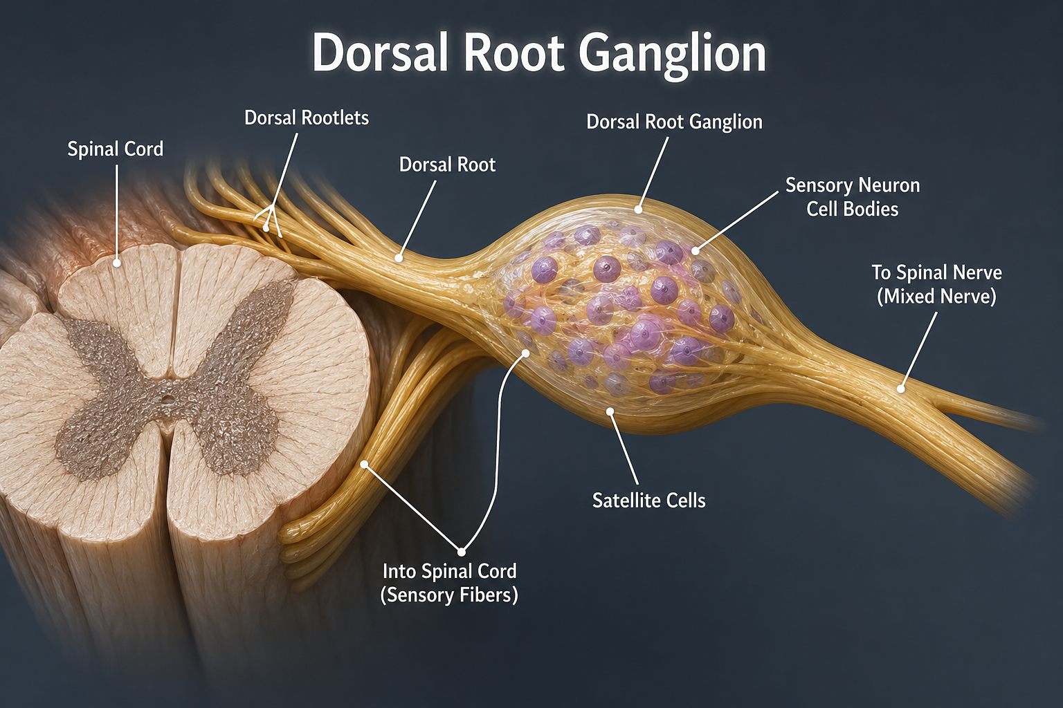
The Dorsal Root Ganglion (DRG), also known as the spinal ganglion, is a swollen enlargement located on the dorsal root of every spinal nerve. It serves as a collection point for the cell bodies of sensory neurons. Although the ganglion processes sensory information, it lies outside the spinal cord and is therefore part of the Peripheral Nervous System (PNS).
🦴 Structure
The dorsal root ganglion contains thousands of sensory neuron cell bodies, each surrounded by satellite cells that provide protection, nourishment, and structural support. Each sensory neuron has a single axon that divides into two branches: one branch extends to sensory receptors in the body, while the other enters the spinal cord through the dorsal root. This unique arrangement allows sensory signals to travel quickly without interruption.
⚙️ Function
The main function of the dorsal root ganglion is to receive and relay sensory information from the body to the spinal cord. It carries signals related to touch, pain, pressure, temperature, vibration, and body position (proprioception). The ganglion also helps maintain the health of sensory neurons by supplying nutrients and supporting their normal function.
🧩 Components
The dorsal root ganglion is mainly composed of sensory neuron cell bodies, satellite glial cells, connective tissue, and blood vessels. Unlike motor ganglia, it does not contain synapses; sensory nerve impulses simply pass through the ganglion before entering the spinal cord.
🔵 Ventral Root (Anterior Root)
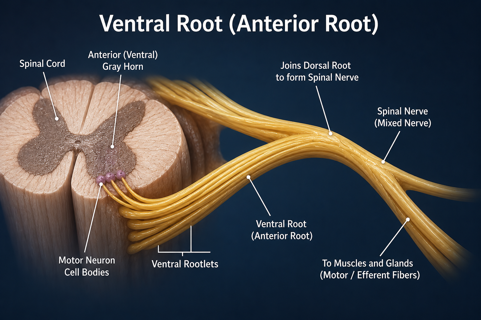
The ventral root, also known as the anterior root, is the motor portion of a spinal nerve. It carries nerve impulses away from the spinal cord to the body's muscles and glands. Unlike the dorsal root, the ventral root contains only motor (efferent) nerve fibers and does not transmit sensory information.
🦴 Structure
The ventral root is formed by numerous small bundles of nerve fibers called ventral rootlets, which arise from the anterior side of the spinal cord. These rootlets merge together to form a single ventral root. The nerve fibers originate from the cell bodies of motor neurons located in the anterior (ventral) gray horn of the spinal cord. Unlike the dorsal root, the ventral root does not contain a ganglion, as the motor neuron cell bodies remain inside the spinal cord.
⚙️ Function
The primary function of the ventral root is to carry motor commands from the central nervous system to skeletal muscles, enabling voluntary movements such as walking, writing, speaking, and lifting objects. In certain spinal regions, it also carries autonomic motor fibers that help regulate involuntary functions of organs and glands.
🧩 Components
The ventral root is composed mainly of motor axons originating from the spinal cord. It is made up of multiple ventral rootlets that unite before joining the dorsal root to form a mixed spinal nerve. Since it contains only motor fibers, all outgoing nerve impulses travel from the spinal cord to their target tissues.
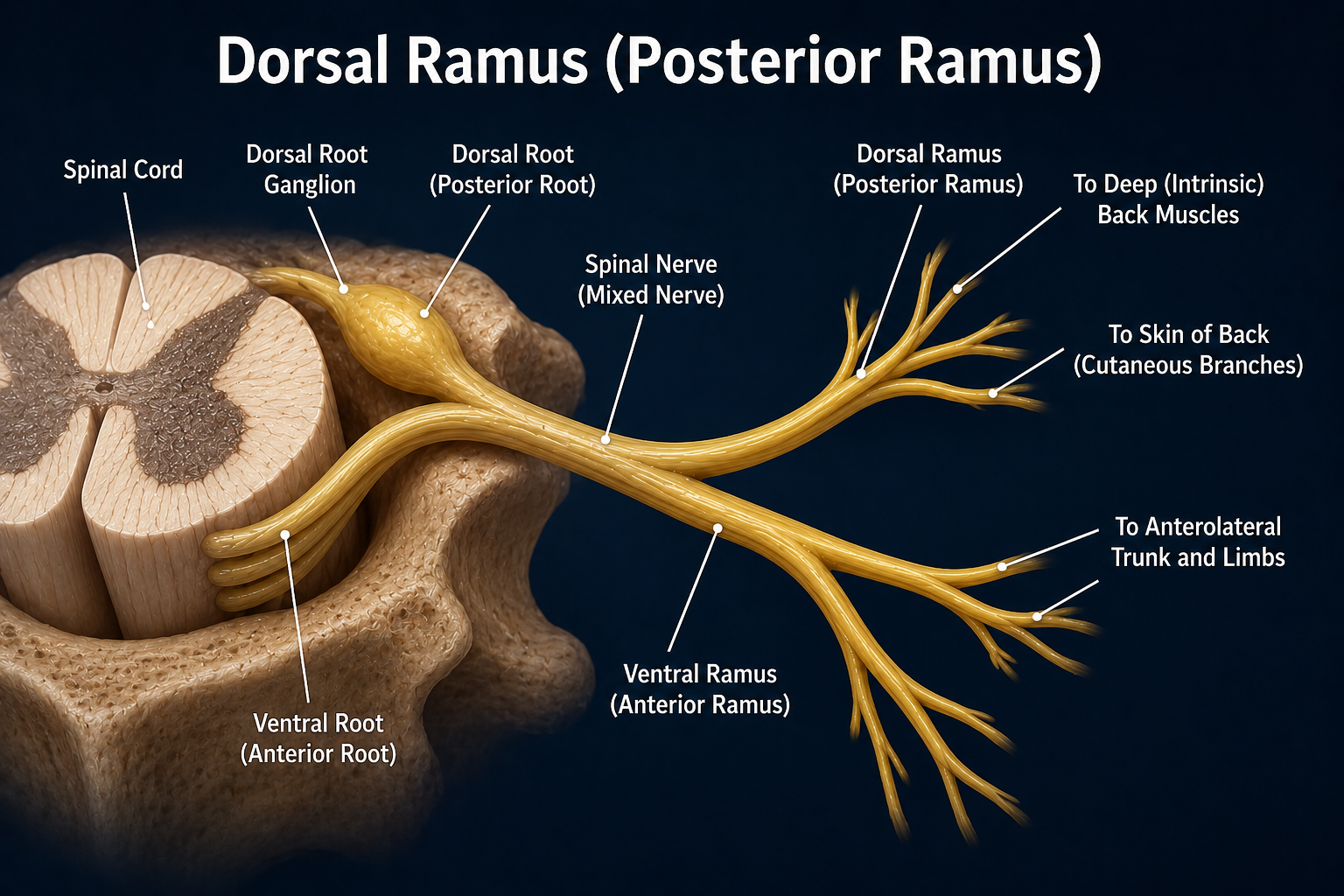
🟡 Dorsal Ramus (Posterior Ramus)
The dorsal ramus, also known as the posterior ramus, is the smaller of the two branches formed after a spinal nerve divides. Despite its smaller size, it is a mixed nerve, containing both sensory and motor fibers. It extends backward from the spinal nerve to supply the structures of the back.
🦴 Structure
Each dorsal ramus divides into medial and lateral branches, which distribute nerves to different regions of the back. These branches contain both sensory and motor fibers and travel through the deep back muscles and overlying skin. Unlike the ventral ramus, the dorsal ramus does not form nerve plexuses.
⚙️ Function
The dorsal ramus supplies the intrinsic (deep) muscles of the back, allowing them to maintain posture and produce movements such as extension, rotation, and side bending of the vertebral column. It also carries sensory information from the skin covering the back and from the vertebral joints and ligaments, helping the brain detect touch, pain, temperature, and body position.
🧩 Further Branches
The dorsal ramus usually divides into two main branches: the medial branch, which supplies structures close to the vertebral column, and the lateral branch, which extends farther outward to innervate the muscles and skin of the back.
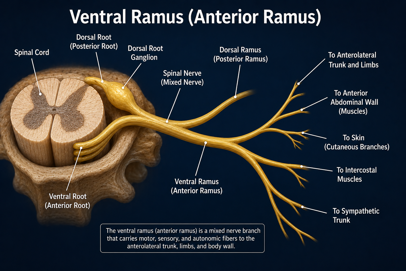
🔴 Ventral Ramus (Anterior Ramus)
The ventral ramus, also known as the anterior ramus, is the larger branch of a spinal nerve. Like the dorsal ramus, it is a mixed nerve, carrying both sensory and motor fibers. After a spinal nerve divides, the ventral ramus travels toward the front and sides of the body to supply the trunk and limbs.
🦴 Structure
The ventral rami are generally much larger than the dorsal rami because they innervate a greater portion of the body. In the cervical, lumbar, and sacral regions, neighboring ventral rami join together to form complex networks called nerve plexuses. However, in the thoracic region, they remain separate and continue as the intercostal nerves that run between the ribs.
⚙️ Function
The ventral ramus supplies the skin and muscles of the front and sides of the trunk, as well as the upper and lower limbs. It carries motor impulses from the spinal cord to muscles, allowing movement, while returning sensory information such as touch, pain, temperature, and pressure back to the central nervous system.
🧩 Further Branches
In most regions of the body, ventral rami combine to form four major nerve plexuses: Cervical Plexus, Brachial Plexus, Lumbar Plexus, and Sacral Plexus. These plexuses give rise to the major nerves that supply the neck, shoulders, arms, pelvis, and legs. In the thoracic region, the ventral rami do not form plexuses but instead become the intercostal nerves.
Mnemonics
"Dogs Guard Very Dangerous Valleys."
"Dragons Grow Very Dangerous Voices."
"Don't Get Very Dirty, Victor!"
Functions of Spinal Nerves
Spinal nerves serve as vital communication pathways between the central nervous system and the rest of the body. As mixed nerves, they carry both sensory and motor information while also contributing to reflex actions and certain autonomic functions. Their coordinated activity enables sensation, movement, and communication between the brain, spinal cord, muscles, and internal organs.
🧠 Transmit Sensory Information
Spinal nerves carry sensory (afferent) impulses from receptors in the skin, muscles, joints, and internal organs to the spinal cord. These signals allow the brain to perceive sensations such as touch, pain, temperature, pressure, vibration, and body position (proprioception).
💪 Control Voluntary Movements
Spinal nerves transmit motor (efferent) impulses from the spinal cord to skeletal muscles. These impulses enable voluntary movements such as walking, writing, speaking, lifting objects, and maintaining posture.
⚡ Conduct Reflex Actions
Spinal nerves play an essential role in spinal reflexes by rapidly transmitting sensory signals to the spinal cord and returning motor responses without requiring immediate brain involvement. This allows quick protective actions, such as withdrawing a hand from a hot surface.
🔄 Connect the CNS and PNS
Spinal nerves serve as communication pathways between the Central Nervous System (CNS) and the Peripheral Nervous System (PNS). They allow continuous two-way communication by carrying sensory information to the spinal cord and motor commands back to the body.
🫀 Regulate Involuntary Functions
Certain spinal nerves also carry autonomic nerve fibers that help regulate involuntary body functions, including heart rate, blood vessel diameter, sweating, digestion, and glandular secretion.
🤸 Supply Different Body Regions
Through their dorsal and ventral rami, spinal nerves provide both sensory and motor innervation to the back, trunk, neck, and limbs. They ensure proper muscle coordination while allowing the skin and deeper tissues to detect external and internal stimuli.
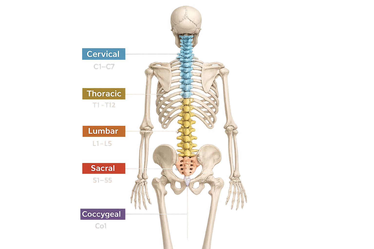
31 Pairs of Spinal Nerves
The human body contains 31 pairs of spinal nerves. Click on any spinal nerve to explore its location and primary functions.
Cervical (8 Pairs)
Thoracic (12 Pairs)
Lumbar (5 Pairs)
Sacral (5 Pairs)
Coccygeal (1 Pair)
Fun Facts About Spinal Nerves
🧠 31 Pairs
Humans have 31 pairs of spinal nerves, making a total of 62 spinal nerves connecting the spinal cord to the body.
⚡ Mixed Nerves
Every spinal nerve is a mixed nerve, carrying both sensory information to the brain and motor commands to muscles.
🏃 Sciatic Nerve
The sciatic nerve, the largest nerve in the body, is formed mainly by the L4–S3 spinal nerves.
📡 Fast Signals
Some spinal nerve fibers conduct electrical impulses at speeds of up to 120 m/s (268 mph).
🦴 8 Cervical Nerves
There are 8 cervical spinal nerves but only 7 cervical vertebrae.
🦶 Achilles Reflex
Doctors test the S1 spinal nerve using the Achilles tendon reflex.
🦵 Knee-Jerk Reflex
The familiar knee-jerk reflex mainly tests the L2–L4 spinal nerves.
🚪 Nerve Exit
Every spinal nerve exits the vertebral column through an opening called an intervertebral foramen.
🔀 Two Branches
Each spinal nerve divides into a dorsal ramus and a ventral ramus after leaving the spine.
💥 Herniated Disc
A slipped (herniated) disc can compress a spinal nerve, causing pain, numbness, or muscle weakness.
💪 Muscle Control
Your muscles cannot move without motor signals carried by spinal nerves.
⚡ Reflex Actions
Spinal nerves allow reflexes to happen before your brain consciously reacts.
🧩 Dorsal Root Ganglion
The dorsal root ganglion contains thousands of sensory neuron cell bodies.
🌡️ Every Sensation
Touch, pain, pressure, vibration, and temperature all travel through spinal nerves.
👶 Before Birth
Spinal nerves begin developing during the first month of embryonic development.
🎯 Dermatomes
Each spinal nerve supplies a specific skin area called a dermatome.
💥 Myotomes
Each spinal nerve also controls specific muscles called a myotome.
❤️ Autonomic Fibers
Spinal nerves carry autonomic fibers that help regulate sweating and blood vessel diameter.
🏋️ Powerful Legs
The lumbar and sacral spinal nerves control the powerful muscles responsible for walking, running, and jumping.
🌍 Communication Highway
The 31 pairs of spinal nerves form the body's main communication network between the spinal cord and the rest of the body.
Your Spinal Cord Ends Before Your Spine Does!
Although your vertebral column extends all the way to your tailbone, the spinal cord actually ends around the L1–L2 vertebrae. Below this point, the remaining spinal nerves continue as a bundle called the Cauda Equina ("Horse's Tail"), allowing doctors to safely perform lumbar punctures without damaging the spinal cord.
C1 Spinal Nerve (First Cervical Nerve)
The C1 spinal nerve, also known as the first cervical nerve, is the uppermost pair of the 31 pairs of spinal nerves. It emerges between the occipital bone of the skull and the first cervical vertebra (atlas). Unlike most spinal nerves, C1 usually contains very few or no sensory fibers, making it primarily a motor nerve.
📍 Location
The C1 spinal nerve exits the vertebral column above the atlas (C1 vertebra). It is the only cervical spinal nerve that emerges above its corresponding vertebra. After leaving the spinal canal, it joins nearby nerves to help form the cervical plexus.
⚙️ Functions
- Supplies motor fibers to several deep muscles of the neck.
- Helps control movements of the head and upper neck.
- Contributes to head flexion, extension, and rotation.
- Participates in the formation of the cervical plexus.
- Assists in maintaining head posture and stability.
🦴 Structures Supplied
- Rectus capitis anterior muscle
- Rectus capitis lateralis muscle
- Longus capitis muscle
- Longus colli muscle
- Several deep suboccipital muscles (through associated branches)
💡 Did You Know?
The C1 spinal nerve is unique because it is primarily motor. In many individuals, it has little or no sensory root, unlike most other spinal nerves, which carry both sensory and motor fibers.
C2 Spinal Nerve (Second Cervical Nerve)
The C2 spinal nerve, also known as the second cervical nerve, is the second pair of cervical spinal nerves. It emerges between the atlas (C1) and the axis (C2). Unlike the C1 nerve, the C2 nerve is a mixed spinal nerve, containing both sensory and motor fibers. It plays an important role in providing sensation to the head and controlling muscles of the upper neck.
📍 Location
The C2 spinal nerve exits the vertebral column between the first (atlas) and second (axis) cervical vertebrae. After leaving the spinal canal, it divides into dorsal and ventral rami, contributing to the cervical plexus and supplying structures of the head and neck.
⚙️ Functions
- Provides sensation to the back of the head and upper neck.
- Supplies motor fibers to several deep neck muscles.
- Assists in head rotation, extension, and neck movements.
- Contributes to the formation of the cervical plexus.
- Supports normal posture and movement of the head.
🦴 Structures Supplied
- Skin of the back of the scalp (via the Greater Occipital Nerve).
- Upper neck skin.
- Suboccipital muscles.
- Semispinalis capitis muscle.
- Deep muscles of the neck.
💡 Did You Know?
The Greater Occipital Nerve, the largest sensory branch of the C2 spinal nerve, supplies most of the skin on the back of the scalp. Irritation or compression of this nerve can cause occipital neuralgia, a condition characterized by severe headaches originating at the base of the skull.
C3 Spinal Nerve (Third Cervical Nerve)
The C3 spinal nerve, also known as the third cervical nerve, is the third pair of cervical spinal nerves. It emerges between the C2 and C3 vertebrae. As a mixed spinal nerve, it carries both sensory and motor fibers. The C3 nerve plays an important role in neck movement, sensation, and breathing by contributing to the phrenic nerve.
📍 Location
The C3 spinal nerve exits the vertebral column through the intervertebral foramen between the second and third cervical vertebrae. After leaving the spinal canal, it divides into dorsal and ventral rami, contributing to the cervical plexus.
⚙️ Functions
- Provides sensation to parts of the neck and the area behind the ear.
- Supplies motor fibers to several muscles of the neck.
- Contributes to head and neck movements.
- Forms part of the phrenic nerve (C3–C5), which controls the diaphragm during breathing.
- Helps maintain posture and stability of the cervical region.
🦴 Structures Supplied
- Skin of the neck.
- Area behind the ear.
- Deep neck muscles.
- Prevertebral muscles.
- Diaphragm (through the phrenic nerve).
💡 Did You Know?
The C3 spinal nerve contributes to the famous anatomical phrase "C3, C4, and C5 keep the diaphragm alive." Together, these spinal nerves form the phrenic nerve, which provides the primary motor supply to the diaphragm—the main muscle responsible for breathing.
C4 Spinal Nerve (Fourth Cervical Nerve)
The C4 spinal nerve, also known as the fourth cervical nerve, is the fourth pair of cervical spinal nerves. It emerges between the C3 and C4 vertebrae. As a mixed spinal nerve, it carries both sensory and motor fibers. The C4 nerve plays a vital role in neck and shoulder function and is one of the major contributors to the phrenic nerve, which controls breathing.
📍 Location
The C4 spinal nerve exits the vertebral column through the intervertebral foramen between the third and fourth cervical vertebrae. After leaving the spinal canal, it divides into dorsal and ventral rami and contributes to the cervical plexus.
⚙️ Functions
- Provides sensation to the lower neck and shoulder region.
- Supplies motor fibers to several muscles of the neck.
- Assists in movements of the neck and shoulders.
- Contributes significantly to the phrenic nerve (C3–C5), which controls the diaphragm.
- Helps maintain normal breathing by supporting diaphragm function.
🦴 Structures Supplied
- Skin of the lower neck.
- Skin over the shoulder (supraclavicular region).
- Levator scapulae muscle.
- Scalene muscles.
- Diaphragm (through the phrenic nerve).
💡 Did You Know?
The C4 spinal nerve provides one of the largest contributions to the phrenic nerve. Damage to the C4 nerve can weaken the diaphragm, making breathing difficult, especially if the neighboring C3 and C5 nerves are also affected.
C5 Spinal Nerve (Fifth Cervical Nerve)
The C5 spinal nerve, also known as the fifth cervical nerve, is the fifth pair of cervical spinal nerves. It emerges between the C4 and C5 vertebrae. As a mixed spinal nerve, it contains both sensory and motor fibers. The C5 nerve is an important contributor to the brachial plexus, which supplies the shoulder and upper limb.
📍 Location
The C5 spinal nerve exits the vertebral column through the intervertebral foramen between the fourth and fifth cervical vertebrae. After leaving the spinal canal, it divides into dorsal and ventral rami. Its ventral ramus joins the brachial plexus, where it helps form several major nerves of the upper limb.
⚙️ Functions
- Provides sensation to the shoulder and the outer (lateral) upper arm.
- Supplies motor fibers to muscles that move the shoulder and elbow.
- Assists in shoulder abduction (lifting the arm sideways).
- Helps flex the elbow through muscles such as the biceps brachii.
- Contributes to the formation of major nerves of the upper limb through the brachial plexus.
🦴 Structures Supplied
- Deltoid muscle.
- Biceps brachii muscle.
- Supraspinatus muscle.
- Infraspinatus muscle.
- Skin over the shoulder and lateral upper arm.
💡 Did You Know?
Doctors often test the C5 spinal nerve by checking the biceps reflex and asking a patient to lift their arm sideways. Weakness in these movements may indicate injury to the C5 nerve or the upper part of the brachial plexus.
C6 Spinal Nerve (Sixth Cervical Nerve)
The C6 spinal nerve, also known as the sixth cervical nerve, is the sixth pair of cervical spinal nerves. It emerges between the C5 and C6 vertebrae. As a mixed spinal nerve, it carries both sensory and motor fibers. The C6 nerve is a major contributor to the brachial plexus, supplying important structures of the shoulder, arm, forearm, and hand.
📍 Location
The C6 spinal nerve exits the vertebral column through the intervertebral foramen between the fifth and sixth cervical vertebrae. After leaving the spinal canal, it divides into dorsal and ventral rami. Its ventral ramus joins the brachial plexus, contributing to several major nerves of the upper limb.
⚙️ Functions
- Provides sensation to the lateral forearm, thumb, and index finger.
- Supplies motor fibers to muscles of the shoulder, arm, and forearm.
- Assists in elbow flexion and wrist extension.
- Helps control movements of the thumb and forearm.
- Contributes to the formation of major upper limb nerves through the brachial plexus.
🦴 Structures Supplied
- Biceps brachii muscle.
- Brachialis muscle.
- Brachioradialis muscle.
- Wrist extensor muscles.
- Skin of the lateral forearm, thumb, and index finger.
💡 Did You Know?
The C6 spinal nerve is commonly tested by checking the brachioradialis reflex. Compression of the C6 nerve root, often caused by a cervical disc herniation, may result in pain, numbness, or tingling that radiates from the neck into the thumb and index finger.
C7 Spinal Nerve (Seventh Cervical Nerve)
The C7 spinal nerve, also known as the seventh cervical nerve, is the seventh pair of cervical spinal nerves. It emerges between the C6 and C7 vertebrae. As a mixed spinal nerve, it carries both sensory and motor fibers. The C7 nerve is one of the largest contributors to the brachial plexus, supplying many muscles and skin areas of the upper limb.
📍 Location
The C7 spinal nerve exits the vertebral column through the intervertebral foramen between the sixth and seventh cervical vertebrae. After leaving the spinal canal, it divides into dorsal and ventral rami. Its ventral ramus joins the brachial plexus, where it contributes to several major nerves that control the arm, forearm, and hand.
⚙️ Functions
- Provides sensation to the back of the forearm and the middle finger.
- Supplies motor fibers to muscles that extend the elbow, wrist, and fingers.
- Helps straighten the elbow through the triceps muscle.
- Assists in wrist and finger extension.
- Contributes to fine motor control of the upper limb through the brachial plexus.
🦴 Structures Supplied
- Triceps brachii muscle.
- Wrist extensor muscles.
- Finger extensor muscles.
- Latissimus dorsi (through associated nerves).
- Skin of the posterior forearm and middle finger.
💡 Did You Know?
The C7 spinal nerve is commonly evaluated by testing the triceps reflex. Compression of the C7 nerve root may cause pain, weakness, or numbness that radiates from the neck down the arm to the middle finger, making it one of the most commonly affected cervical nerve roots.
C8 Spinal Nerve (Eighth Cervical Nerve)
The C8 spinal nerve, also known as the eighth cervical nerve, is the final pair of cervical spinal nerves. Unlike the other cervical nerves, it emerges between the C7 vertebra and the T1 vertebra. As a mixed spinal nerve, it carries both sensory and motor fibers. The C8 nerve is an important contributor to the brachial plexus, supplying muscles and skin of the forearm and hand.
📍 Location
The C8 spinal nerve exits the vertebral column through the intervertebral foramen between the seventh cervical (C7) and first thoracic (T1) vertebrae. It is the only cervical spinal nerve that emerges below the C7 vertebra. After exiting the spinal canal, it divides into dorsal and ventral rami, with the ventral ramus becoming part of the brachial plexus.
⚙️ Functions
- Provides sensation to the ring finger, little finger, and the inner side of the forearm.
- Supplies motor fibers to many muscles of the hand and forearm.
- Assists in finger flexion and extension.
- Controls fine finger movements such as gripping, writing, and typing.
- Contributes to the formation of major nerves of the upper limb through the brachial plexus.
🦴 Structures Supplied
- Flexor digitorum profundus muscle.
- Flexor digitorum superficialis muscle.
- Intrinsic muscles of the hand.
- Finger flexor muscles.
- Skin of the little finger, ring finger, and medial forearm.
💡 Did You Know?
The C8 spinal nerve is unique because there are 8 cervical spinal nerves but only 7 cervical vertebrae. C8 exits between the C7 and T1 vertebrae. Injury to the C8 nerve may cause weakness in hand grip, difficulty performing fine finger movements, and numbness in the little finger and ring finger.
T1 Spinal Nerve (First Thoracic Nerve)
The T1 spinal nerve, also known as the first thoracic nerve, is the first pair of thoracic spinal nerves. It emerges between the T1 and T2 vertebrae. As a mixed spinal nerve, it carries both sensory and motor fibers. The T1 nerve is unique because, unlike the other thoracic nerves, it contributes to the brachial plexus, helping supply the upper limb in addition to the upper chest.
📍 Location
The T1 spinal nerve exits the vertebral column through the intervertebral foramen between the first and second thoracic vertebrae. After emerging from the spinal canal, it divides into dorsal and ventral rami. The ventral ramus joins the brachial plexus, while the dorsal ramus supplies the muscles and skin of the upper back.
⚙️ Functions
- Provides sensation to the upper back and the inner side of the upper arm.
- Supplies motor fibers to muscles of the upper back and shoulder region.
- Contributes to the brachial plexus, helping control muscles of the forearm and hand.
- Supports posture and movement of the upper thoracic region.
- Carries sympathetic nerve fibers to the head, neck, and upper limb.
🦴 Structures Supplied
- Skin of the upper back.
- Skin of the medial (inner) upper arm.
- Deep muscles of the upper back.
- Intrinsic muscles of the hand (through the brachial plexus).
- Sympathetic fibers supplying the upper limb.
💡 Did You Know?
The T1 spinal nerve is the only thoracic spinal nerve that normally becomes part of the brachial plexus. This is why injuries to the T1 nerve can affect the small muscles of the hand and weaken grip strength, despite being a thoracic nerve.
T2 Spinal Nerve (Second Thoracic Nerve)
The T2 spinal nerve, also known as the second thoracic nerve, is the second pair of thoracic spinal nerves. It emerges between the T2 and T3 vertebrae. As a mixed spinal nerve, it carries both sensory and motor fibers. The T2 nerve mainly supplies the upper chest, upper back, and the armpit (axillary) region.
📍 Location
The T2 spinal nerve exits the vertebral column through the intervertebral foramen between the second and third thoracic vertebrae. After emerging from the spinal canal, it divides into dorsal and ventral rami. The ventral ramus continues as the second intercostal nerve, while a large lateral branch, known as the Intercostobrachial Nerve, supplies the skin of the armpit and upper medial arm.
⚙️ Functions
- Provides sensation to the upper chest and upper back.
- Supplies sensation to the armpit (axilla) and medial upper arm.
- Supplies motor fibers to the second intercostal muscles.
- Assists in movements of the rib cage during breathing.
- Carries sympathetic nerve fibers to the thoracic wall.
🦴 Structures Supplied
- Second intercostal muscles.
- Skin of the upper chest.
- Skin of the upper back.
- Skin of the axilla (armpit).
- Skin of the medial upper arm (via the Intercostobrachial Nerve).
💡 Did You Know?
The Intercostobrachial Nerve, a branch of the T2 spinal nerve, is often affected during breast or axillary surgery. Injury to this nerve may cause numbness or tingling in the inner upper arm after the procedure.
T3 Spinal Nerve (Third Thoracic Nerve)
The T3 spinal nerve, also known as the third thoracic nerve, is the third pair of thoracic spinal nerves. It emerges between the T3 and T4 vertebrae. As a mixed spinal nerve, it carries both sensory and motor fibers. The T3 nerve primarily supplies the upper chest, upper back, and the muscles located between the ribs.
📍 Location
The T3 spinal nerve exits the vertebral column through the intervertebral foramen between the third and fourth thoracic vertebrae. It divides into dorsal and ventral rami after leaving the spinal canal. The ventral ramus continues as the third intercostal nerve, while the dorsal ramus supplies the muscles and skin of the upper back.
⚙️ Functions
- Provides sensation to the upper chest and upper back.
- Supplies motor fibers to the third intercostal muscles.
- Helps stabilize and move the rib cage during breathing.
- Supports posture by supplying muscles of the upper back.
- Carries sympathetic nerve fibers to structures within the thoracic region.
🦴 Structures Supplied
- Third intercostal muscles.
- Skin of the upper chest.
- Skin of the upper back.
- Deep muscles of the upper thoracic region.
- Blood vessels and sweat glands of the T3 dermatome through sympathetic fibers.
💡 Did You Know?
The T3 spinal nerve contributes to the third intercostal nerve, which plays an important role in expanding and contracting the chest wall during breathing. Damage to this nerve may cause pain or numbness along the third rib and nearby chest wall.
T4 Spinal Nerve (Fourth Thoracic Nerve)
The T4 spinal nerve, also known as the fourth thoracic nerve, is the fourth pair of thoracic spinal nerves. It emerges between the T4 and T5 vertebrae. As a mixed spinal nerve, it carries both sensory and motor fibers. The T4 nerve supplies the chest wall and intercostal muscles, playing an important role in breathing and chest sensation.
📍 Location
The T4 spinal nerve exits the vertebral column through the intervertebral foramen between the fourth and fifth thoracic vertebrae. It divides into dorsal and ventral rami. The ventral ramus continues as the fourth intercostal nerve, while the dorsal ramus supplies the muscles and skin of the upper back.
⚙️ Functions
- Provides sensation to the chest wall at the level of the nipples.
- Supplies motor fibers to the fourth intercostal muscles.
- Helps expand and contract the rib cage during breathing.
- Supports posture by supplying muscles of the upper back.
- Carries sympathetic nerve fibers to the skin, blood vessels, and sweat glands of the chest.
🦴 Structures Supplied
- Fourth intercostal muscles.
- Skin of the anterior and lateral chest wall.
- Skin of the upper back.
- Deep muscles of the thoracic region.
- Blood vessels and sweat glands within the T4 dermatome.
💡 Did You Know?
The T4 dermatome is commonly used as an anatomical landmark because it corresponds approximately to the nipple line in most adults. Medical professionals often use this landmark during neurological examinations to assess sensation and identify possible spinal nerve injuries.
T5 Spinal Nerve (Fifth Thoracic Nerve)
The T5 spinal nerve, also known as the fifth thoracic nerve, is the fifth pair of thoracic spinal nerves. It emerges between the T5 and T6 vertebrae. As a mixed spinal nerve, it carries both sensory and motor fibers. The T5 nerve supplies the middle chest wall and the intercostal muscles, contributing to breathing and sensation in the thoracic region.
📍 Location
The T5 spinal nerve exits the vertebral column through the intervertebral foramen between the fifth and sixth thoracic vertebrae. After leaving the spinal canal, it divides into dorsal and ventral rami. The ventral ramus continues as the fifth intercostal nerve, while the dorsal ramus supplies the muscles and skin of the mid-back.
⚙️ Functions
- Provides sensation to the middle region of the chest and upper back.
- Supplies motor fibers to the fifth intercostal muscles.
- Helps expand and stabilize the rib cage during breathing.
- Supports movements and posture of the thoracic wall.
- Carries sympathetic nerve fibers to the skin, blood vessels, and sweat glands of the T5 dermatome.
🦴 Structures Supplied
- Fifth intercostal muscles.
- Skin of the middle chest wall.
- Skin of the mid-back.
- Deep muscles of the thoracic region.
- Blood vessels and sweat glands within the T5 dermatome.
💡 Did You Know?
The T5 spinal nerve forms the fifth intercostal nerve, which runs beneath the fifth rib alongside an artery and vein. Together, these structures form the intercostal neurovascular bundle, an important landmark that surgeons carefully avoid during chest procedures.
T6 Spinal Nerve (Sixth Thoracic Nerve)
The T6 spinal nerve, also known as the sixth thoracic nerve, is the sixth pair of thoracic spinal nerves. It emerges between the T6 and T7 vertebrae. As a mixed spinal nerve, it carries both sensory and motor fibers. The T6 nerve supplies the lower chest wall, intercostal muscles, and part of the upper abdominal region, contributing to breathing, posture, and trunk sensation.
📍 Location
The T6 spinal nerve exits the vertebral column through the intervertebral foramen between the sixth and seventh thoracic vertebrae. After leaving the spinal canal, it divides into dorsal and ventral rami. The ventral ramus continues as the sixth intercostal nerve, while the dorsal ramus supplies the muscles and skin of the middle back.
⚙️ Functions
- Provides sensation to the lower chest and upper abdominal wall.
- Supplies motor fibers to the sixth intercostal muscles.
- Assists in expanding and stabilizing the rib cage during breathing.
- Supports movements and posture of the thoracic wall.
- Carries sympathetic nerve fibers to the skin, blood vessels, and sweat glands of the T6 dermatome.
🦴 Structures Supplied
- Sixth intercostal muscles.
- Skin of the lower chest wall.
- Skin of the upper abdominal region.
- Deep muscles of the middle back.
- Blood vessels and sweat glands within the T6 dermatome.
💡 Did You Know?
The T6 spinal nerve marks the transition between the middle and lower chest. Through the sixth intercostal nerve, it helps coordinate the movement of the rib cage during breathing while also providing sensation to the upper abdominal wall.
T7 Spinal Nerve (Seventh Thoracic Nerve)
The T7 spinal nerve, also known as the seventh thoracic nerve, is the seventh pair of thoracic spinal nerves. It emerges between the T7 and T8 vertebrae. As a mixed spinal nerve, it carries both sensory and motor fibers. Unlike the upper thoracic nerves, the T7 nerve extends beyond the rib cage to supply both the lower chest and the upper abdominal wall.
📍 Location
The T7 spinal nerve exits the vertebral column through the intervertebral foramen between the seventh and eighth thoracic vertebrae. After emerging from the spinal canal, it divides into dorsal and ventral rami. The ventral ramus continues as the seventh intercostal nerve, supplying the lower chest before continuing into the upper abdominal wall.
⚙️ Functions
- Provides sensation to the lower chest and upper abdomen.
- Supplies motor fibers to the seventh intercostal muscles.
- Assists in movements of the rib cage during breathing.
- Helps strengthen the upper abdominal wall.
- Carries sympathetic nerve fibers to the skin, blood vessels, and sweat glands of the T7 dermatome.
🦴 Structures Supplied
- Seventh intercostal muscles.
- Upper abdominal muscles.
- Skin of the lower chest.
- Skin of the upper abdominal wall.
- Deep muscles of the thoracic region.
💡 Did You Know?
The T7 spinal nerve is one of the first thoracic nerves whose intercostal branch continues into the abdominal wall. This allows it to help control the upper abdominal muscles, which are important for posture, coughing, and forceful exhalation.
T8 Spinal Nerve (Eighth Thoracic Nerve)
The T8 spinal nerve, also known as the eighth thoracic nerve, is the eighth pair of thoracic spinal nerves. It emerges between the T8 and T9 vertebrae. As a mixed spinal nerve, it carries both sensory and motor fibers. The T8 nerve supplies the lower chest wall and continues into the upper abdominal wall, helping in trunk movement and sensation.
📍 Location
The T8 spinal nerve exits the vertebral column through the intervertebral foramen between the eighth and ninth thoracic vertebrae. After leaving the spinal canal, it divides into dorsal and ventral rami. The ventral ramus continues as the eighth intercostal nerve, supplying the lower chest before passing into the upper abdominal wall.
⚙️ Functions
- Provides sensation to the lower chest and upper abdominal wall.
- Supplies motor fibers to the eighth intercostal muscles.
- Assists in breathing by helping move the lower ribs.
- Helps in movements of the upper abdominal muscles.
- Carries sympathetic nerve fibers to the skin, blood vessels, and sweat glands of the T8 dermatome.
🦴 Structures Supplied
- Eighth intercostal muscles.
- Upper abdominal muscles.
- Skin of the lower chest wall.
- Skin of the upper abdominal wall.
- Deep muscles of the thoracic region.
💡 Did You Know?
The T8 spinal nerve is one of the thoracic nerves that extends beyond the rib cage to supply the abdominal wall. Along with the lower thoracic nerves, it helps control the muscles used during coughing, sneezing, forceful exhalation, and maintaining abdominal pressure.
T9 Spinal Nerve (Ninth Thoracic Nerve)
The T9 spinal nerve, also known as the ninth thoracic nerve, is the ninth pair of thoracic spinal nerves. It emerges between the T9 and T10 vertebrae. As a mixed spinal nerve, it carries both sensory and motor fibers. The T9 nerve primarily supplies the lower chest and upper abdominal wall, contributing to breathing, posture, and abdominal muscle movements.
📍 Location
The T9 spinal nerve exits the vertebral column through the intervertebral foramen between the ninth and tenth thoracic vertebrae. It divides into dorsal and ventral rami after leaving the spinal canal. The ventral ramus continues as the ninth intercostal nerve, extending into the upper abdominal wall, while the dorsal ramus supplies the muscles and skin of the back.
⚙️ Functions
- Provides sensation to the lower chest and upper abdominal wall.
- Supplies motor fibers to the ninth intercostal muscles.
- Assists in expanding and stabilizing the rib cage during breathing.
- Helps control the upper abdominal muscles during trunk movements.
- Carries sympathetic nerve fibers to the skin, blood vessels, and sweat glands of the T9 dermatome.
🦴 Structures Supplied
- Ninth intercostal muscles.
- Upper abdominal muscles.
- Skin of the lower chest.
- Skin of the upper abdominal wall.
- Deep muscles of the thoracic region.
💡 Did You Know?
The T9 spinal nerve contributes to the nerves of the anterior abdominal wall. Along with the surrounding thoracic nerves, it helps coordinate movements such as bending, twisting the trunk, coughing, sneezing, and maintaining abdominal pressure.
T10 Spinal Nerve (Tenth Thoracic Nerve)
The T10 spinal nerve, also known as the tenth thoracic nerve, is the tenth pair of thoracic spinal nerves. It emerges between the T10 and T11 vertebrae. As a mixed spinal nerve, it carries both sensory and motor fibers. The T10 nerve supplies the lower abdominal wall and is best known for its dermatome, which corresponds to the umbilicus (navel).
📍 Location
The T10 spinal nerve exits the vertebral column through the intervertebral foramen between the tenth and eleventh thoracic vertebrae. After emerging from the spinal canal, it divides into dorsal and ventral rami. The ventral ramus continues forward as the tenth intercostal nerve, supplying the lower abdominal wall, while the dorsal ramus innervates the muscles and skin of the back.
⚙️ Functions
- Provides sensation to the lower abdominal wall.
- Supplies motor fibers to the tenth intercostal muscles.
- Helps control the abdominal muscles during movement and posture.
- Assists in breathing by supporting movements of the lower rib cage.
- Carries sympathetic nerve fibers to the skin, blood vessels, and sweat glands of the T10 dermatome.
🦴 Structures Supplied
- Tenth intercostal muscles.
- Upper portions of the abdominal muscles.
- Skin surrounding the umbilical (navel) region.
- Deep muscles of the lower thoracic region.
- Blood vessels and sweat glands within the T10 dermatome.
💡 Did You Know?
The T10 dermatome corresponds approximately to the umbilicus (belly button). During neurological examinations, doctors often test sensation around the navel to assess the function of the T10 spinal nerve and detect possible spinal cord or nerve root injuries.
T11 Spinal Nerve (Eleventh Thoracic Nerve)
The T11 spinal nerve, also known as the eleventh thoracic nerve, is the eleventh pair of thoracic spinal nerves. It emerges between the T11 and T12 vertebrae. As a mixed spinal nerve, it carries both sensory and motor fibers. The T11 nerve primarily supplies the lower abdominal wall and contributes to movements of the trunk and abdominal muscles.
📍 Location
The T11 spinal nerve exits the vertebral column through the intervertebral foramen between the eleventh and twelfth thoracic vertebrae. After leaving the spinal canal, it divides into dorsal and ventral rami. The ventral ramus continues as the eleventh intercostal nerve, extending into the lower abdominal wall, while the dorsal ramus supplies the muscles and skin of the lower back.
⚙️ Functions
- Provides sensation to the lower abdominal wall.
- Supplies motor fibers to the eleventh intercostal muscles.
- Helps control the abdominal muscles during bending and twisting movements.
- Assists in forceful breathing, coughing, and sneezing.
- Carries sympathetic nerve fibers to the skin, blood vessels, and sweat glands of the T11 dermatome.
🦴 Structures Supplied
- Eleventh intercostal muscles.
- Lower abdominal muscles.
- Skin of the lower abdominal wall.
- Deep muscles of the lower thoracic region.
- Blood vessels and sweat glands within the T11 dermatome.
💡 Did You Know?
The T11 spinal nerve helps supply the lower abdominal muscles, which play an important role in maintaining posture, stabilizing the trunk, and increasing abdominal pressure during activities such as lifting heavy objects, coughing, and sneezing.
T12 Spinal Nerve (Twelfth Thoracic Nerve)
The T12 spinal nerve, also known as the twelfth thoracic nerve, is the final pair of thoracic spinal nerves. It emerges below the T12 vertebra and is a mixed spinal nerve, carrying both sensory and motor fibers. Unlike the other thoracic nerves, its ventral ramus is called the Subcostal Nerve because it runs beneath the twelfth rib instead of between two ribs.
📍 Location
The T12 spinal nerve exits the vertebral column through the intervertebral foramen between the twelfth thoracic vertebra and the first lumbar vertebra (L1). After emerging from the spinal canal, it divides into dorsal and ventral rami. The ventral ramus continues as the Subcostal Nerve, supplying the lower abdominal wall and part of the hip region.
⚙️ Functions
- Provides sensation to the lower abdomen and upper hip region.
- Supplies motor fibers to the abdominal wall muscles.
- Helps stabilize the trunk during movement.
- Assists in forceful breathing, coughing, and sneezing.
- Carries sympathetic nerve fibers to the skin, blood vessels, and sweat glands of the lower trunk.
🦴 Structures Supplied
- Subcostal muscle.
- External oblique muscle.
- Internal oblique muscle.
- Transversus abdominis muscle.
- Skin of the lower abdominal wall and upper hip region.
💡 Did You Know?
The T12 spinal nerve is unique because its ventral ramus forms the Subcostal Nerve, which travels below the twelfth rib. Unlike the other thoracic nerves, it is not an intercostal nerve, making it an important link between the thoracic and lumbar regions.
L1 Spinal Nerve (First Lumbar Nerve)
The L1 spinal nerve, also known as the first lumbar nerve, is the first pair of lumbar spinal nerves. It emerges between the L1 and L2 vertebrae. As a mixed spinal nerve, it carries both sensory and motor fibers. The L1 nerve supplies the lower abdominal wall, groin region, and upper thigh, while also contributing to movements of the abdominal muscles.
📍 Location
The L1 spinal nerve exits the vertebral column through the intervertebral foramen between the first and second lumbar vertebrae. After leaving the spinal canal, it divides into dorsal and ventral rami. The ventral ramus contributes to the lumbar plexus and gives rise to the iliohypogastric and ilioinguinal nerves.
⚙️ Functions
- Provides sensation to the lower abdomen, groin, and upper thigh.
- Supplies motor fibers to portions of the abdominal wall muscles.
- Helps support posture and trunk stability.
- Contributes to movements such as bending and twisting the trunk.
- Carries sympathetic nerve fibers to the lower abdominal region.
🦴 Structures Supplied
- External oblique muscle.
- Internal oblique muscle.
- Transversus abdominis muscle.
- Skin of the lower abdomen.
- Skin of the groin and upper medial thigh.
💡 Did You Know?
The L1 spinal nerve is the first spinal nerve to form part of the lumbar plexus. Through its branches—the iliohypogastric and ilioinguinal nerves—it provides sensation to the groin and lower abdominal wall and helps strengthen the abdominal muscles.
L2 Spinal Nerve (Second Lumbar Nerve)
The L2 spinal nerve, also known as the second lumbar nerve, is the second pair of lumbar spinal nerves. It emerges between the L2 and L3 vertebrae. As a mixed spinal nerve, it carries both sensory and motor fibers. The L2 nerve contributes significantly to the lumbar plexus, supplying the lower abdomen, hip, groin, and upper thigh while helping control movements of the hip and thigh.
📍 Location
The L2 spinal nerve exits the vertebral column through the intervertebral foramen between the second and third lumbar vertebrae. After leaving the spinal canal, it divides into dorsal and ventral rami. The ventral ramus becomes part of the lumbar plexus, contributing fibers to the genitofemoral, lateral femoral cutaneous, femoral, and obturator nerves.
⚙️ Functions
- Provides sensation to the groin, upper thigh, and hip region.
- Supplies motor fibers to muscles involved in hip flexion.
- Contributes to movements of the thigh and hip joint.
- Supports posture and balance during standing and walking.
- Carries sympathetic nerve fibers to structures of the lower limb.
🦴 Structures Supplied
- Psoas major muscle (partially).
- Iliacus muscle (through the femoral nerve).
- Skin of the upper anterior thigh.
- Skin of the groin region.
- Hip joint through branches of the lumbar plexus.
💡 Did You Know?
The L2 spinal nerve plays an important role in forming the femoral and obturator nerves, two of the largest nerves of the lumbar plexus. These nerves are essential for walking because they help control the muscles that flex the hip and move the thigh.
L3 Spinal Nerve (Third Lumbar Nerve)
The L3 spinal nerve, also known as the third lumbar nerve, is the third pair of lumbar spinal nerves. It emerges between the L3 and L4 vertebrae. As a mixed spinal nerve, it carries both sensory and motor fibers. The L3 nerve is a major contributor to the lumbar plexus, supplying the front and inner thigh while helping control movements of the hip and knee.
📍 Location
The L3 spinal nerve exits the vertebral column through the intervertebral foramen between the third and fourth lumbar vertebrae. After leaving the spinal canal, it divides into dorsal and ventral rami. The ventral ramus contributes to the femoral, obturator, and lateral femoral cutaneous nerves of the lumbar plexus.
⚙️ Functions
- Provides sensation to the front and inner side of the thigh.
- Supplies motor fibers to muscles that flex the hip.
- Helps extend the knee through the femoral nerve.
- Contributes to thigh adduction through the obturator nerve.
- Supports standing, walking, and climbing stairs.
🦴 Structures Supplied
- Quadriceps femoris muscles (through the femoral nerve).
- Adductor muscles of the thigh (through the obturator nerve).
- Psoas major muscle.
- Skin of the anterior and medial thigh.
- Hip and knee joints through branches of the lumbar plexus.
💡 Did You Know?
The L3 spinal nerve is important for the patellar (knee-jerk) reflex. Together with the L2 and L4 spinal nerves, it helps produce this reflex, which doctors commonly test during neurological examinations to assess nerve function.
L4 Spinal Nerve (Fourth Lumbar Nerve)
The L4 spinal nerve, also known as the fourth lumbar nerve, is the fourth pair of lumbar spinal nerves. It emerges between the L4 and L5 vertebrae. As a mixed spinal nerve, it carries both sensory and motor fibers. The L4 nerve is a major contributor to both the lumbar plexus and the lumbosacral trunk, making it an important connection between the lumbar and sacral plexuses.
📍 Location
The L4 spinal nerve exits the vertebral column through the intervertebral foramen between the fourth and fifth lumbar vertebrae. After leaving the spinal canal, it divides into dorsal and ventral rami. The ventral ramus contributes to the femoral and obturator nerves, while part of it joins the lumbosacral trunk, which continues into the sacral plexus.
⚙️ Functions
- Provides sensation to the front of the knee, medial leg, and ankle.
- Supplies motor fibers to muscles that extend the knee.
- Helps control ankle dorsiflexion (lifting the foot upward).
- Contributes to thigh adduction through the obturator nerve.
- Forms part of the lumbosacral trunk, linking the lumbar and sacral plexuses.
🦴 Structures Supplied
- Quadriceps femoris muscles.
- Tibialis anterior muscle (partly through the lumbosacral trunk).
- Adductor muscles of the thigh.
- Skin of the medial leg and ankle.
- Knee and ankle joints through branches of the lumbar plexus.
💡 Did You Know?
The L4 spinal nerve is one of the primary nerves tested during the patellar (knee-jerk) reflex. It is also unique because part of it forms the lumbosacral trunk, creating the important connection between the lumbar and sacral plexuses that supply most of the lower limb.
L5 Spinal Nerve (Fifth Lumbar Nerve)
The L5 spinal nerve, also known as the fifth lumbar nerve, is the final pair of lumbar spinal nerves. It emerges between the L5 and S1 vertebrae. As a mixed spinal nerve, it carries both sensory and motor fibers. The L5 nerve is a major contributor to the lumbosacral plexus, supplying the hip, leg, foot, and toes. It plays a vital role in walking, running, and maintaining balance.
📍 Location
The L5 spinal nerve exits the vertebral column through the intervertebral foramen between the fifth lumbar and first sacral vertebrae. After leaving the spinal canal, it divides into dorsal and ventral rami. The ventral ramus joins the lumbosacral trunk, becoming an important part of the sacral plexus, which gives rise to the large nerves of the lower limb, including the sciatic nerve.
⚙️ Functions
- Provides sensation to the outer leg, top of the foot, and great toe.
- Supplies motor fibers to muscles that lift the foot and extend the toes.
- Helps control hip abduction and knee stabilization during walking.
- Contributes to movements of the ankle and foot.
- Forms part of the sciatic nerve through the lumbosacral plexus.
🦴 Structures Supplied
- Tibialis anterior muscle.
- Extensor hallucis longus muscle.
- Gluteus medius and gluteus minimus muscles.
- Skin of the lateral leg, dorsum (top) of the foot, and great toe.
- Ankle and foot joints through branches of the lumbosacral plexus.
💡 Did You Know?
The L5 spinal nerve is one of the most commonly compressed spinal nerves due to a herniated disc at the L4–L5 level. Injury to this nerve may cause foot drop, making it difficult to lift the front of the foot while walking, and can also lead to numbness over the great toe.
S1 Spinal Nerve (First Sacral Nerve)
The S1 spinal nerve, also known as the first sacral nerve, is the first pair of sacral spinal nerves. It exits the vertebral canal through the first sacral foramen (S1). As a mixed spinal nerve, it carries both sensory and motor fibers. The S1 nerve is a major contributor to the sacral plexus and forms an important part of the sciatic nerve, supplying the buttocks, back of the leg, foot, and ankle.
📍 Location
The S1 spinal nerve leaves the spinal canal through the first sacral foramen. After exiting, it divides into dorsal and ventral rami. The ventral ramus joins the sacral plexus, contributing to the sciatic, superior gluteal, and inferior gluteal nerves, while the dorsal ramus supplies muscles and skin over the back of the sacrum.
⚙️ Functions
- Provides sensation to the buttock, back of the leg, outer foot, and little toe.
- Supplies motor fibers to muscles responsible for ankle plantarflexion (pointing the foot downward).
- Helps control movements of the hip, knee, ankle, and foot.
- Contributes to walking, running, jumping, and standing on tiptoes.
- Forms a major part of the sciatic nerve.
🦴 Structures Supplied
- Gastrocnemius muscle.
- Soleus muscle.
- Gluteus maximus (through the inferior gluteal nerve).
- Skin of the posterior leg and lateral foot.
- Ankle and foot joints through branches of the sciatic nerve.
💡 Did You Know?
The S1 spinal nerve is tested using the Achilles (ankle-jerk) reflex. Doctors tap the Achilles tendon to evaluate S1 nerve function. Damage to this nerve may cause weakness when standing on tiptoes, reduced ankle reflexes, and pain that travels down the back of the leg—a common feature of sciatica.
S2 Spinal Nerve (Second Sacral Nerve)
The S2 spinal nerve, also known as the second sacral nerve, is the second pair of sacral spinal nerves. It exits the vertebral canal through the second sacral foramen (S2). As a mixed spinal nerve, it carries both sensory and motor fibers. The S2 nerve contributes to the sacral plexus, supplying the pelvis, buttocks, back of the thigh, leg, foot, and pelvic organs.
📍 Location
The S2 spinal nerve exits through the second sacral foramen. It divides into dorsal and ventral rami after leaving the spinal canal. The ventral ramus joins the sacral plexus, contributing fibers to the sciatic, pudendal, and posterior femoral cutaneous nerves, while the dorsal ramus supplies muscles and skin over the sacral region.
⚙️ Functions
- Provides sensation to the buttocks, back of the thigh, leg, foot, and perineum.
- Supplies motor fibers to muscles of the lower limb and pelvic floor.
- Helps control movements of the ankle, foot, and toes.
- Contributes to bladder, bowel, and sexual functions through the sacral plexus.
- Forms part of the sciatic and pudendal nerves.
🦴 Structures Supplied
- Hamstring muscles (through the sciatic nerve).
- Intrinsic muscles of the foot.
- Pelvic floor muscles.
- Skin of the posterior thigh, leg, and perineum.
- Bladder, bowel, and reproductive organs through autonomic pathways.
💡 Did You Know?
The S2 spinal nerve is one of the main nerves involved in controlling the bladder, bowel, and sexual organs. Together with S3 and S4, it provides essential nerve supply to the pelvic organs, making it crucial for normal urinary, digestive, and reproductive functions.
S3 Spinal Nerve (Third Sacral Nerve)
The S3 spinal nerve, also known as the third sacral nerve, is the third pair of sacral spinal nerves. It exits the vertebral canal through the third sacral foramen (S3). As a mixed spinal nerve, it carries both sensory and motor fibers. The S3 nerve contributes to the sacral plexus, supplying the pelvis, pelvic floor, buttocks, lower limb, and pelvic organs.
📍 Location
The S3 spinal nerve exits through the third sacral foramen. After emerging from the sacral canal, it divides into dorsal and ventral rami. The ventral ramus contributes to the sacral plexus and pudendal nerve, while the dorsal ramus supplies the muscles and skin over the sacral region.
⚙️ Functions
- Provides sensation to the buttocks, perineum, and pelvic region.
- Supplies motor fibers to the pelvic floor muscles.
- Helps control bladder and bowel function.
- Contributes to sexual function through the pudendal nerve.
- Supports pelvic organ stability and continence.
🦴 Structures Supplied
- Pelvic floor muscles.
- External urethral sphincter.
- External anal sphincter.
- Skin of the perineum and surrounding pelvic region.
- Pelvic organs through branches of the sacral plexus.
💡 Did You Know?
The S3 spinal nerve, together with S2 and S4, forms part of the pudendal nerve, which is responsible for voluntary control of urination and defecation. Damage to these nerves may lead to bladder or bowel incontinence and reduced sensation in the pelvic region.
S4 Spinal Nerve (Fourth Sacral Nerve)
The S4 spinal nerve, also known as the fourth sacral nerve, is the fourth pair of sacral spinal nerves. It exits the vertebral canal through the fourth sacral foramen (S4). As a mixed spinal nerve, it carries both sensory and motor fibers. The S4 nerve is primarily responsible for supplying the pelvic floor, perineum, and pelvic organs, making it essential for urinary, bowel, and sexual functions.
📍 Location
The S4 spinal nerve exits through the fourth sacral foramen. After leaving the sacral canal, it divides into dorsal and ventral rami. The ventral ramus contributes to the sacral plexus, pudendal nerve, and pelvic splanchnic nerves, while the dorsal ramus supplies the muscles and skin over the sacral region.
⚙️ Functions
- Provides sensation to the perineum and area surrounding the anus.
- Supplies motor fibers to the pelvic floor muscles.
- Helps control bladder and bowel emptying.
- Contributes to normal sexual function.
- Supports the pelvic organs by maintaining muscle tone.
🦴 Structures Supplied
- Levator ani muscle.
- Coccygeus muscle.
- External anal sphincter.
- Skin of the perineal region.
- Bladder, rectum, and reproductive organs through pelvic autonomic nerves.
💡 Did You Know?
The S4 spinal nerve contributes to the pelvic splanchnic nerves (S2–S4), which carry parasympathetic fibers to the bladder, rectum, and reproductive organs. A common medical mnemonic is "S2, S3, S4 keep the pee, poop, and penis off the floor", highlighting their role in pelvic organ function and continence.
S5 Spinal Nerve (Fifth Sacral Nerve)
The S5 spinal nerve, also known as the fifth sacral nerve, is the final pair of sacral spinal nerves. It exits the vertebral canal through the sacral hiatus or the fifth sacral foramen. As a mixed spinal nerve, it carries both sensory and motor fibers. The S5 nerve mainly supplies the skin around the tailbone and anus while contributing to the muscles of the pelvic floor and the normal function of pelvic organs.
📍 Location
The S5 spinal nerve leaves the sacral canal through the sacral hiatus or the fifth sacral foramen. After exiting, it divides into dorsal and ventral rami. The ventral ramus joins the sacral plexus and contributes to the coccygeal plexus, supplying structures around the pelvic floor and coccyx.
⚙️ Functions
- Provides sensation to the skin around the coccyx (tailbone) and anus.
- Supplies motor fibers to parts of the pelvic floor muscles.
- Helps support the bladder, bowel, and reproductive organs.
- Contributes to normal bowel and bladder control.
- Forms part of the coccygeal plexus.
🦴 Structures Supplied
- Levator ani muscle (partially).
- Coccygeus muscle.
- Skin over the coccyx and around the anus.
- Pelvic floor structures.
- Pelvic organs through the coccygeal plexus.
💡 Did You Know?
Although the S5 spinal nerve is one of the smallest spinal nerves, it plays an important role in supporting the pelvic floor. Together with S4 and the coccygeal nerve, it helps form the coccygeal plexus, which supplies the skin over the tailbone and contributes to pelvic stability.
Co1 Spinal Nerve (First Coccygeal Nerve)
The Co1 spinal nerve, also known as the first coccygeal nerve, is the only pair of coccygeal spinal nerves and the 31st pair of spinal nerves. It exits the vertebral canal through the sacral hiatus, just below the coccyx (tailbone). As a mixed spinal nerve, it carries both sensory and motor fibers. Although it is the smallest spinal nerve, it contributes to the coccygeal plexus, supplying the skin around the coccyx and supporting the pelvic floor.
📍 Location
The Co1 spinal nerve emerges from the end of the spinal canal through the sacral hiatus, below the fifth sacral nerve (S5). It joins with branches of the S4 and S5 spinal nerves to form the coccygeal plexus. From there, it supplies the skin and muscles surrounding the coccyx and pelvic floor.
⚙️ Functions
- Provides sensation to the skin over the coccyx (tailbone).
- Supplies motor fibers to portions of the pelvic floor muscles.
- Helps support the pelvic organs.
- Contributes to the coccygeal plexus.
- Assists in maintaining stability of the pelvic floor.
🦴 Structures Supplied
- Skin over the coccyx (tailbone).
- Coccygeus muscle.
- Levator ani muscle (partially).
- Pelvic floor connective tissues.
- Structures supplied through the coccygeal plexus.
💡 Did You Know?
The Co1 spinal nerve is the smallest and final spinal nerve in the human body. It joins with the S4 and S5 spinal nerves to form the coccygeal plexus, which supplies the skin around the tailbone. Although tiny, it completes the 31 pairs of spinal nerves that connect the spinal cord to the rest of the body.