Interactive Spinal Cord Labelled Illustration
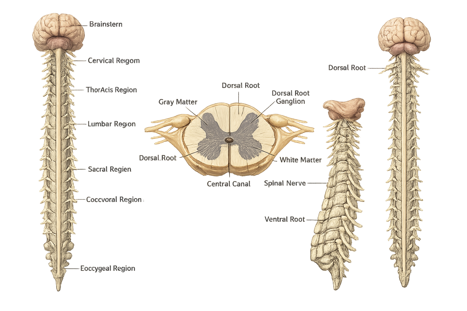
The spinal cord is a long, thin, cylindrical structure made of nervous tissue that forms part of the central nervous system (CNS) along with the brain. It is a continuation of the brainstem, beginning at the medulla oblongata, and extends downward through the vertebral column (backbone) to the lumbar region, where it tapers into the cauda equina. The spinal cord lies inside the spinal canal and is protected by the vertebral column, covered by three protective membranes called the meninges, and surrounded by cerebrospinal fluid (CSF), which cushions it from injury. At its center is a narrow central canal that also contains cerebrospinal fluid. In adults, the spinal cord is approximately 43–45 cm long and extends from the base of the skull to the lower back. It serves as the main communication pathway between the brain and the rest of the body, carrying nerve signals through its network of neurons.
The spinal cord is much more than just a connection between the brain and the rest of the body. It serves as a major communication pathway, carrying sensory and motor information while also controlling rapid reflex actions. By working closely with the brain and peripheral nerves, the spinal cord helps coordinate movement, maintain posture, and regulate several important body functions.
The spinal cord serves as the main communication pathway between the brain and the rest of the body by transmitting nerve impulses in both directions.
It carries sensory impulses such as touch, pain, temperature, and pressure from sensory receptors to the brain for interpretation.
The spinal cord transmits motor signals from the brain to muscles, enabling voluntary movements such as walking, writing, and lifting objects.
It acts as the reflex center for rapid automatic responses, such as the knee-jerk reflex and withdrawing the hand from a hot object.
The spinal cord links the brain with the Peripheral Nervous System (PNS), allowing continuous communication throughout the body.
It helps coordinate muscle activity by transmitting signals between the brain and muscles, ensuring smooth and controlled movements.
The spinal cord produces immediate protective reflexes that help prevent injuries before the brain consciously processes the situation.
It works with the brain and muscles to maintain body posture and support balance during standing, walking, and other activities.
Motor neurons within the spinal cord stimulate skeletal muscles, allowing precise control of voluntary muscle contractions.
The spinal cord processes simple neural pathways by integrating sensory input with motor output during reflex actions.
Ascending tracts carry sensory information to the brain, while descending tracts carry motor instructions from the brain to different parts of the body.
The spinal cord contains pathways that help regulate autonomic functions such as blood vessel diameter, sweating, bladder control, and bowel function.
The human Spinal cord is the main pathway connecting your brain to the rest of your body. It is commonly divided into five regions based on the spinal nerves that arise from it.
The cervical region is the uppermost part of the spinal cord, located within the neck. It contains 8 cervical spinal nerves (C1–C8) that supply the head, neck, shoulders, arms, and diaphragm. It is essential for breathing, upper-limb movement, and sensation.
The thoracic region lies in the chest and gives rise to 12 thoracic spinal nerves (T1–T12). It supplies the chest muscles, ribs, upper abdomen, and internal organs, while also helping maintain posture and trunk stability.
The lumbar region is located in the lower back and contains 5 lumbar spinal nerves (L1–L5). It controls the hips, thighs, legs, and feet, making it important for standing, walking, and lower-body movement.
The sacral region is found near the pelvis and consists of 5 sacral spinal nerves (S1–S5). It supplies the pelvic organs, buttocks, legs, and feet, and helps regulate bladder, bowel, and reproductive functions.
The coccygeal region is the smallest and lowest part of the spinal cord. It contains one coccygeal spinal nerve (Co1) that supplies the skin around the tailbone and contributes to sensory function in this area.
The cervical region is the uppermost part of the spinal cord, located in the neck. It extends from the base of the skull to the seventh cervical vertebra (C7). This region contains 8 pairs of cervical spinal nerves (C1–C8) that connect the brain with the head, neck, shoulders, arms, and hands.
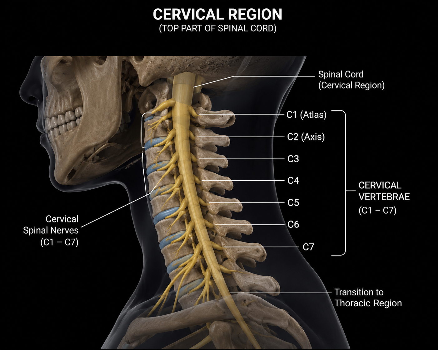
The cervical enlargement is the widest part of the cervical spinal cord. It contains a large number of motor and sensory neurons that supply the shoulders, arms, forearms, and hands. This enlargement allows precise control of upper-limb movements.
The cervical region gives rise to eight pairs of cervical spinal nerves (C1–C8). These nerves transmit sensory information to the brain and carry motor commands to the muscles of the head, neck, shoulders, diaphragm, and upper limbs.
The gray matter forms the butterfly-shaped center of the cervical spinal cord. It contains the cell bodies of neurons and serves as the main site for processing nerve signals and coordinating reflex actions.
The white matter surrounds the gray matter and consists of myelinated nerve fibers. It contains ascending tracts that carry sensory information to the brain and descending tracts that carry motor signals to the body.
The central canal is a narrow channel running through the center of the cervical spinal cord. It contains cerebrospinal fluid (CSF), which nourishes nervous tissue and helps protect the spinal cord from injury.
The cervical spinal cord is enclosed by three protective membranes called the meninges—the dura mater, arachnoid mater, and pia mater. These layers protect the spinal cord and contain the cerebrospinal fluid.
The cervical region is an essential part of the spinal cord that connects the brain with the upper parts of the body. It plays an important role in controlling movement, receiving sensory information, and supporting vital activities such as breathing. Its network of cervical spinal nerves allows proper communication between the nervous system and the head, neck, and upper limbs.
The thoracic region is the second region of the spinal cord, located in the chest. It extends through the thoracic vertebrae (T1–T12) and contains 12 pairs of thoracic spinal nerves. This region connects the brain with the chest, upper back, and parts of the abdomen.
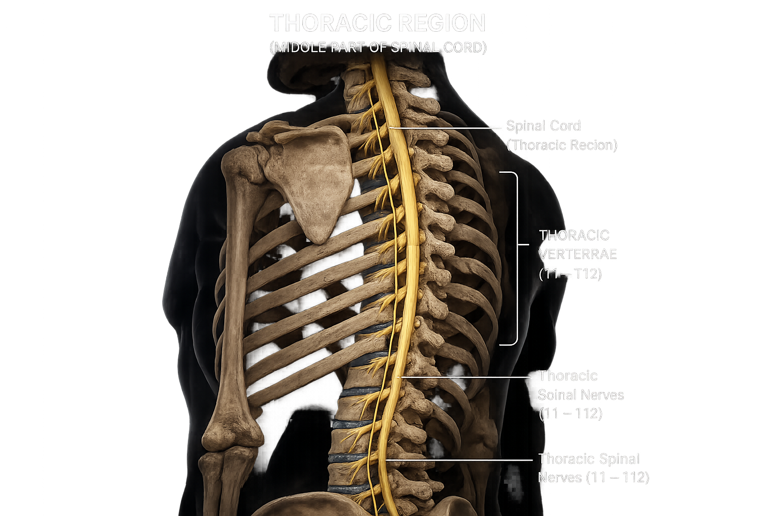
The thoracic vertebrae consist of 12 vertebrae (T1–T12) located in the middle portion of the vertebral column. They are larger than the cervical vertebrae but smaller than the lumbar vertebrae. Each thoracic vertebra has facets for rib attachment, allowing them to form the posterior part of the rib cage. These vertebrae provide strength, protect the thoracic spinal cord, and help support the upper body while limiting excessive movement to maintain stability.
The thoracic spinal cord is the section of the spinal cord that extends through the thoracic vertebral canal. It acts as a major communication pathway between the brain and the chest, back, and upper abdominal regions. It carries ascending sensory impulses to the brain and descending motor commands to muscles. It also participates in several reflex activities and contains pathways of the autonomic nervous system that regulate many involuntary body functions.
There are 12 pairs of thoracic spinal nerves, one pair emerging from each thoracic vertebra. These mixed nerves contain both sensory and motor fibers. They transmit sensory information such as touch, pain, pressure, and temperature from the chest and upper abdomen to the brain, while motor fibers control the muscles of the chest wall, upper back, and abdominal region.
The anterior branches of most thoracic spinal nerves continue as the intercostal nerves, which run between adjacent ribs. These nerves supply the intercostal muscles responsible for expanding and contracting the rib cage during breathing. They also provide sensation to the skin of the chest, ribs, and upper abdominal wall, making them essential for both movement and sensory perception in the thoracic region.
The thoracic region is the longest section of the spinal cord. It serves as an important pathway between the brain and the chest, upper back, and abdomen, helping to coordinate movement, transmit sensory information, and support various automatic body functions.
The lumbar region is the third region of the spinal cord, located in the lower back. It extends through the lumbar vertebrae (L1–L5) and contains 5 pairs of lumbar spinal nerves. This region connects the brain with the lower back, hips, legs, and feet.
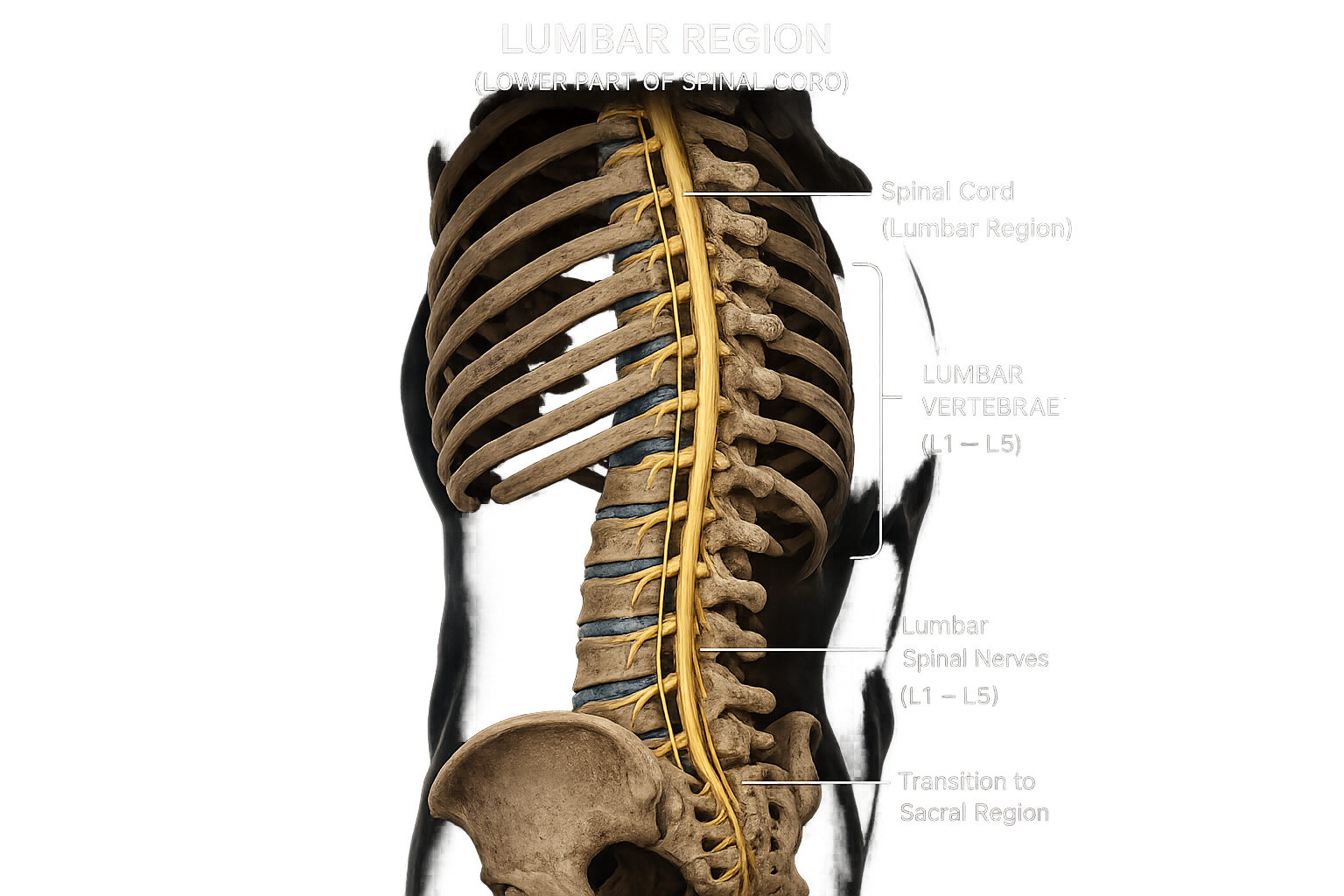
The lumbar vertebrae consist of five large vertebrae (L1–L5) located in the lower back. They are the largest and strongest vertebrae in the vertebral column because they support most of the body's weight. These vertebrae provide stability, allow movements such as bending, twisting, and lifting, and protect the lumbar portion of the spinal cord and nerve roots.
The lumbar region of the spinal cord is responsible for transmitting sensory and motor impulses between the brain and the lower limbs. It plays an essential role in controlling movements of the hips, legs, knees, ankles, and feet, while also participating in reflex actions involving the lower body.
The five pairs of lumbar spinal nerves emerge from the lumbar region of the spinal cord. These mixed nerves carry sensory information from the lower abdomen, hips, thighs, legs, and feet to the brain, while motor fibers control the muscles responsible for standing, walking, and other lower limb movements.
The cauda equina, meaning "horse's tail", is a bundle of spinal nerve roots that begins below the end of the spinal cord. Since the spinal cord ends around the L1–L2 vertebral level, these nerve roots continue downward inside the vertebral canal before exiting through the lumbar, sacral, and coccygeal openings. They provide nerve supply to the legs, bladder, bowel, and pelvic organs.
The lumbar region is an important part of the spinal cord that connects the brain with the lower body. It plays a vital role in controlling the hips, legs, and feet, allowing smooth movement, transmitting sensory information, and supporting everyday activities such as walking, running, and maintaining balance.
The sacral region is the fourth region of the spinal cord, located in the pelvic area. It extends through the sacral vertebrae (S1–S5) and contains 5 pairs of sacral spinal nerves. This region connects the brain with the pelvis, buttocks, legs, feet, and several pelvic organs.
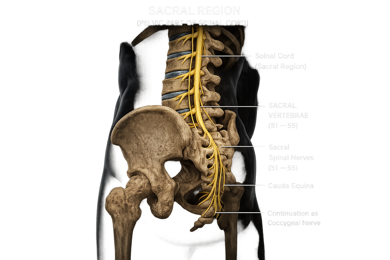
The sacral region consists of five spinal cord segments (S1–S5). These segments give rise to the sacral spinal nerves, which carry both sensory and motor impulses between the spinal cord and the lower parts of the body.
The sacral nerves emerge from the sacral spinal cord and supply the pelvis, buttocks, thighs, legs, feet, and pelvic organs. They control voluntary muscle movements and transmit sensations such as touch, pain, and temperature.
The sacral plexus is a network of nerves formed by the lower lumbar and sacral spinal nerves. It gives rise to important nerves, including the sciatic nerve, which supplies most of the lower limb.
The filum terminale is a thin strand of connective tissue extending downward from the end of the spinal cord. It anchors the spinal cord to the coccyx (tailbone), helping keep it stable within the vertebral canal.
The sacral canal is the continuation of the vertebral canal through the sacrum. It protects the sacral nerve roots as they travel toward the lower body and pelvic organs.
The sacral hiatus is an opening at the lower end of the sacral canal. It allows passage of the filum terminale and is commonly used as an access point for caudal epidural anesthesia.
The sacral region is an important part of the spinal cord that connects the brain with the pelvic region and lower limbs. It plays a vital role in controlling movement, transmitting sensory information, and regulating the functions of important pelvic organs, helping the body perform everyday activities efficiently.
The coccygeal region is the lowest and smallest region of the spinal cord. It is located near the tailbone (coccyx) and contains 1 pair of coccygeal spinal nerves (Co1). This region connects the brain with the area around the coccyx and contributes to the sensory and motor functions of the lower end of the body.
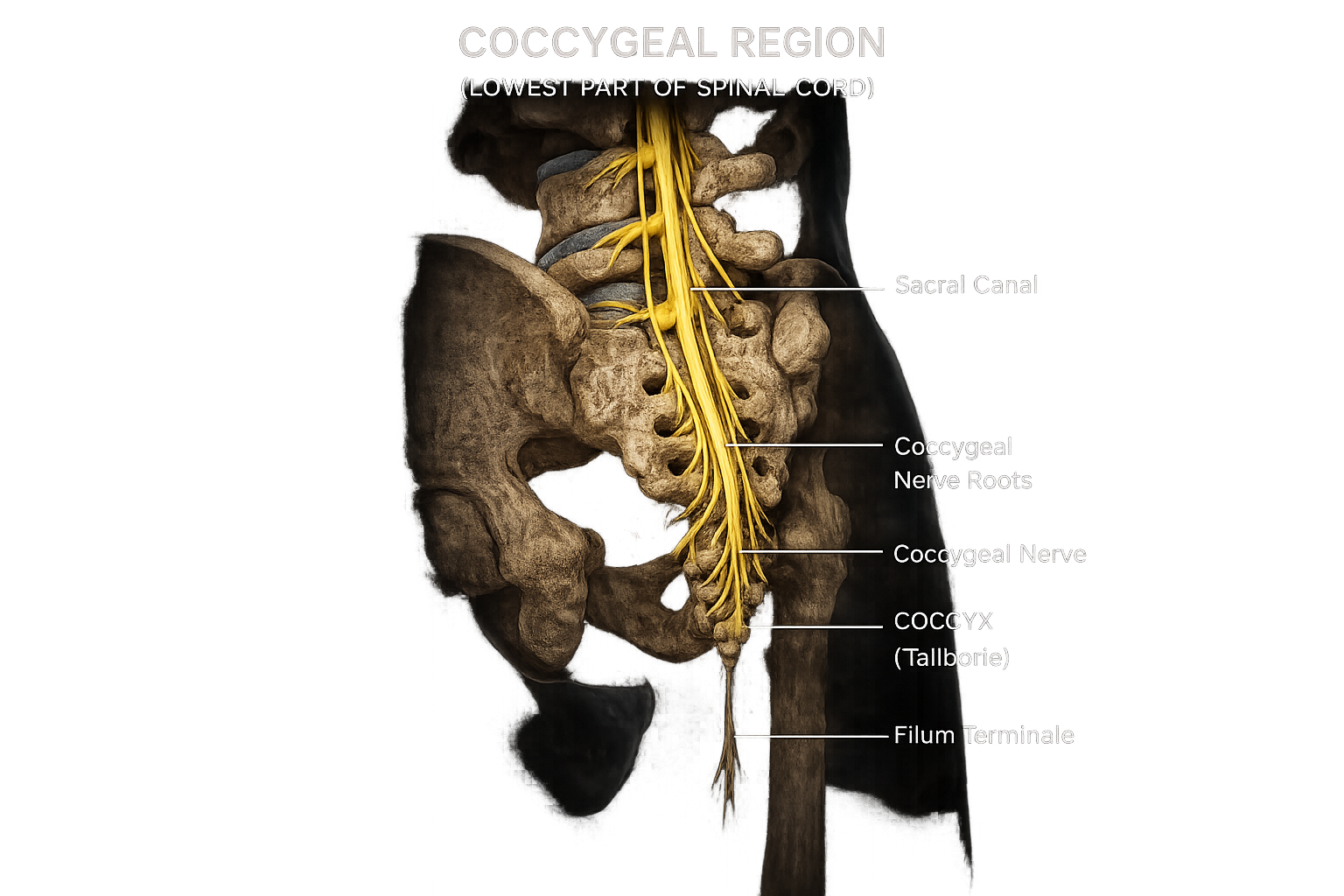
The coccyx, commonly known as the tailbone, is the terminal part of the vertebral column. It is formed by the fusion of three to five small vertebrae, usually four. Although it has limited movement, it provides an attachment site for ligaments, tendons, and pelvic floor muscles, helping support the pelvic organs.
The coccygeal nerve (Co1) is the smallest spinal nerve. It provides sensory innervation to the skin around the coccyx and contributes to the coccygeal plexus, which supplies structures in the pelvic floor.
The coccygeal nerve roots arise from the terminal end of the spinal cord and join with sacral nerves to form the coccygeal plexus. They carry both sensory and motor nerve impulses to the tissues surrounding the tailbone and pelvic floor.
The filum terminale is a thin, fibrous strand of connective tissue that extends from the end of the spinal cord to the coccyx. Its primary function is to anchor and stabilize the spinal cord within the vertebral canal, preventing excessive movement.
The sacral canal extends downward into the coccygeal region and contains the filum terminale and lower nerve roots. It serves as a protected passageway for these structures before they reach the coccyx.
The coccygeal plexus is a small network of nerves formed by the coccygeal nerve and portions of the fourth and fifth sacral nerves. It supplies the skin over the coccyx and contributes to the muscles and tissues of the pelvic floor.
The coccygeal region is the smallest and lowest part of the spinal cord. Although it is small, it plays an important role in transmitting sensory and motor signals to the area around the tailbone and helps support the normal function of the pelvic region.
Here are some fascinating facts about the Spinal cord!
The adult spinal cord is only about 43–45 cm (17–18 inches) long.
Nerve impulses can travel through the spinal cord at speeds of up to 120 meters per second.
The spinal cord is protected by the vertebral column, meninges, and cerebrospinal fluid (CSF).
The spinal cord actually floats in cerebrospinal fluid, which cushions it from shocks.
The spinal cord ends near the L1–L2 vertebrae, not at the bottom of the backbone.
The bundle of nerves below the spinal cord is called the cauda equina, meaning "horse's tail."
Many reflex actions are controlled by the spinal cord without waiting for the brain.
The spinal cord is the body's main communication highway between the brain and the rest of the body.
It gives rise to 31 pairs of spinal nerves that connect the CNS with the entire body.
Doctors often test the knee-jerk reflex to check spinal cord function.
The spinal cord contains Central Pattern Generators (CPGs) that help produce walking movements.
Unlike many tissues, the spinal cord has very limited ability to regenerate after injury.
Together with the brain, the spinal cord forms the Central Nervous System (CNS).
The spinal cord is enclosed inside the vertebral (spinal) canal for protection.
It helps regulate bladder function, bowel control, sweating, and blood vessel diameter.
The spinal cord is divided into five regions: cervical, thoracic, lumbar, sacral, and coccygeal.
The sciatic nerve, the largest nerve in the human body, originates from the lower spinal cord.
During fetal development, the spinal cord initially extends the entire length of the vertebral column.
The spinal cord carries millions of electrical signals every second between the brain and body.
Without the spinal cord, the brain could not communicate with most parts of the body.