Interactive Lungs Labelled Illustration
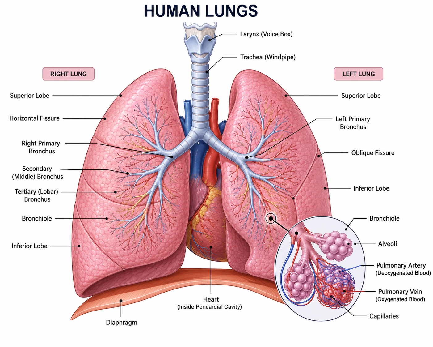
🫁 External Anatomy of the Lungs
The lungs are a pair of soft, spongy organs located inside the thoracic cavity. Although they appear similar, each lung has unique features that help fit around the heart and other organs. Understanding the external anatomy of the lungs helps explain how they efficiently support breathing and gas exchange.
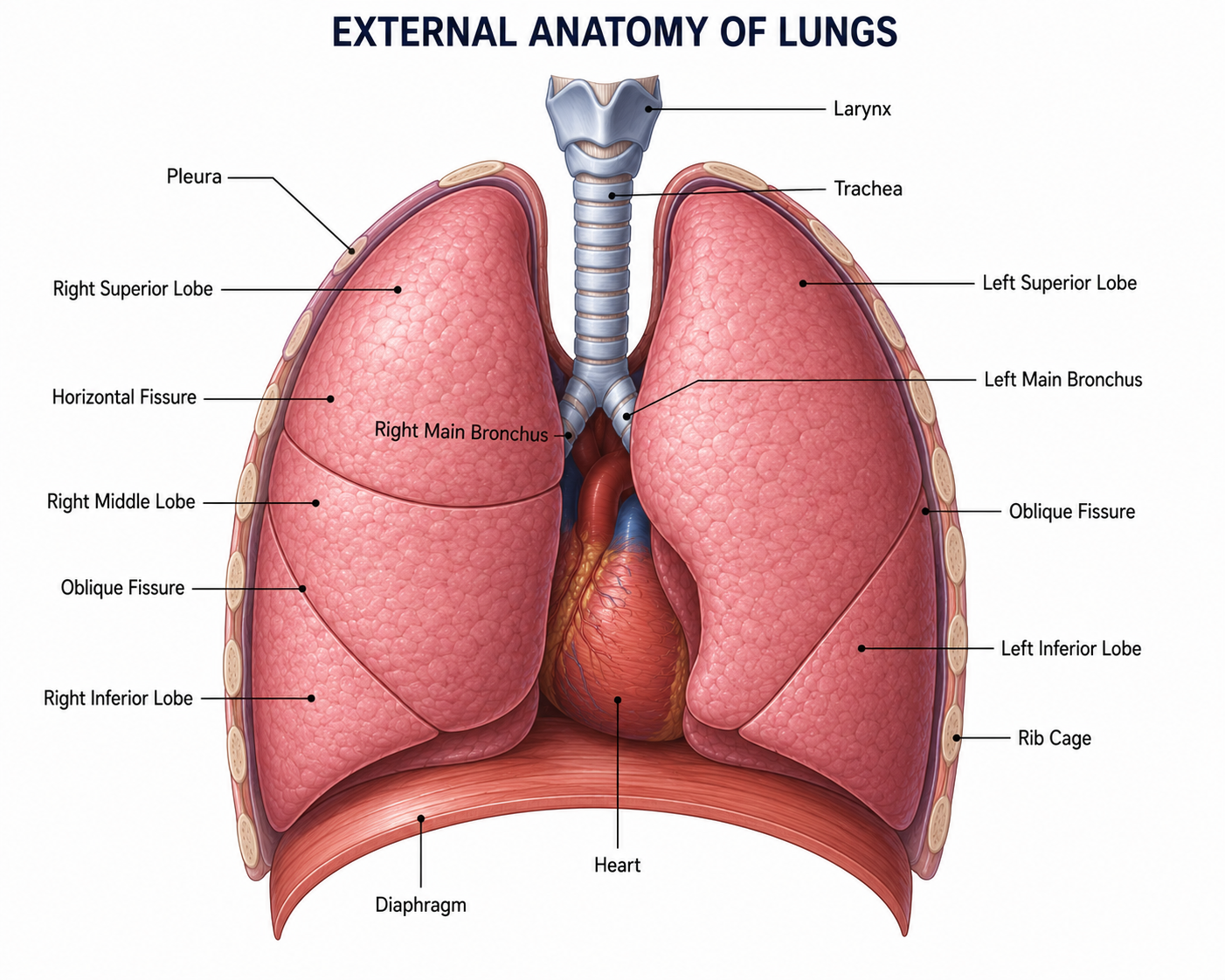
Right Lung
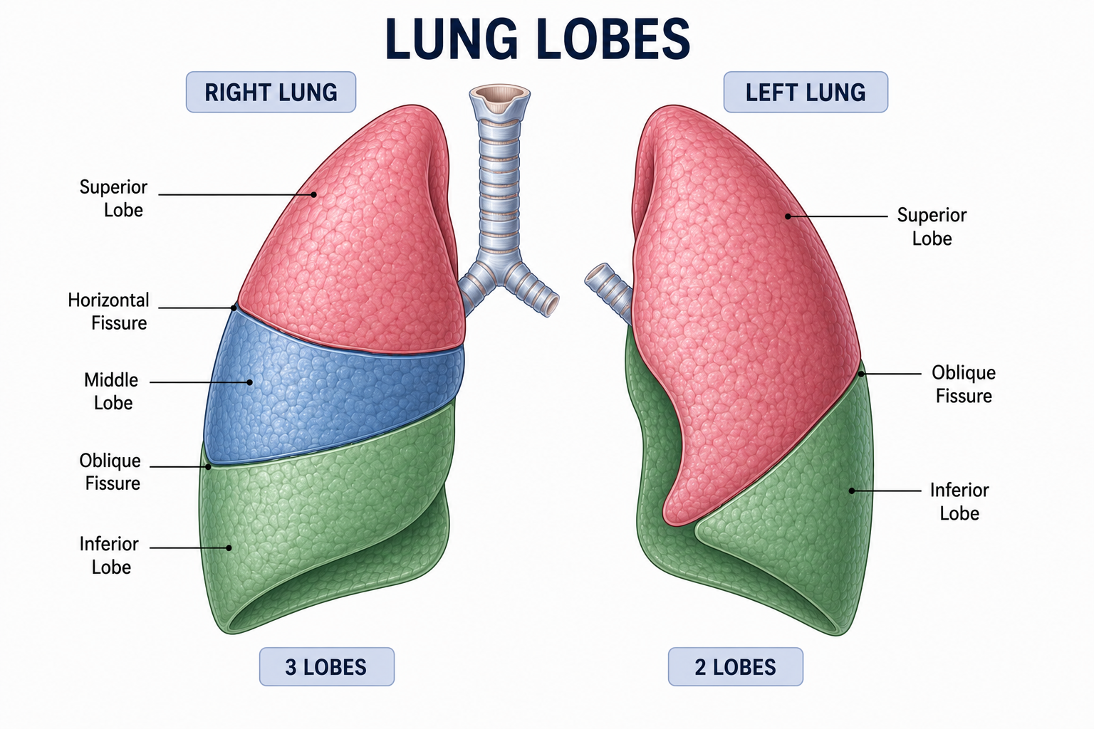
The right lung is the larger of the two lungs. It consists of three lobes (superior, middle, and inferior) and is shorter and wider because the liver lies beneath it.
Left Lung
The left lung is slightly smaller and contains only two lobes. It has a special indentation called the cardiac notch, which provides space for the heart.
Apex
The apex is the pointed upper end of each lung. It extends slightly above the collarbone and is the highest part of the lungs.
Base
The base is the broad lower surface of the lungs. It rests directly on the diaphragm, the main muscle responsible for breathing.
Hilum
The hilum is the central region on the inner surface of each lung where the bronchi, blood vessels, lymphatic vessels, and nerves enter and leave the lungs.
Cardiac Notch
The cardiac notch is a curved depression found only on the left lung. It allows the heart to fit comfortably within the chest cavity without compressing the lung.
🔬 Internal Anatomy of the Lungs
Inside the lungs is an intricate network of air passages that becomes progressively smaller as air travels deeper into the respiratory system. This branching network, known as the bronchial tree, ensures that oxygen reaches every tiny air sac called an alveolus, where gas exchange takes place. Each structure has a specific role in transporting, distributing, and exchanging gases efficiently.
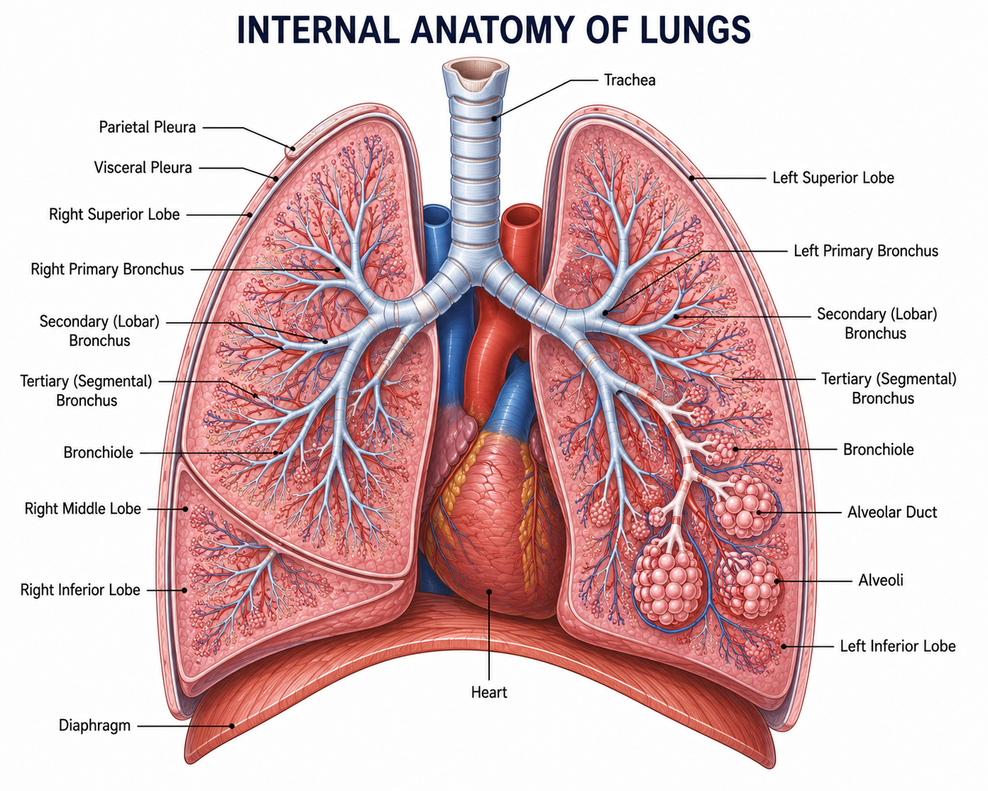
Bronchi
The right and left bronchi are the two main branches of the trachea. They carry air into each lung and divide into smaller branches that distribute air throughout the lungs.
Bronchioles
Bronchioles are tiny branches of the bronchi. Their smooth muscles regulate airflow by widening or narrowing the airways according to the body's oxygen requirements.
Alveolar Ducts
These narrow passages connect the respiratory bronchioles to the alveolar sacs, allowing air to reach the microscopic gas exchange surfaces.
Alveolar Sacs
Alveolar sacs are clusters of tiny alveoli grouped together like bunches of grapes. They maximize the surface available for oxygen and carbon dioxide exchange.
Alveoli
The alveoli are microscopic air sacs where oxygen enters the blood and carbon dioxide leaves the blood through diffusion.
Capillary Network
Every alveolus is surrounded by numerous blood capillaries. These tiny vessels collect oxygen and transport it throughout the body while returning carbon dioxide to the lungs.
⚖️ Right Lung vs Left Lung
Although both lungs perform the same function, they are not identical. The right lung is larger and consists of three lobes, while the left lung is slightly smaller with two lobes because it shares space with the heart. These differences allow both lungs to fit perfectly within the thoracic cavity.
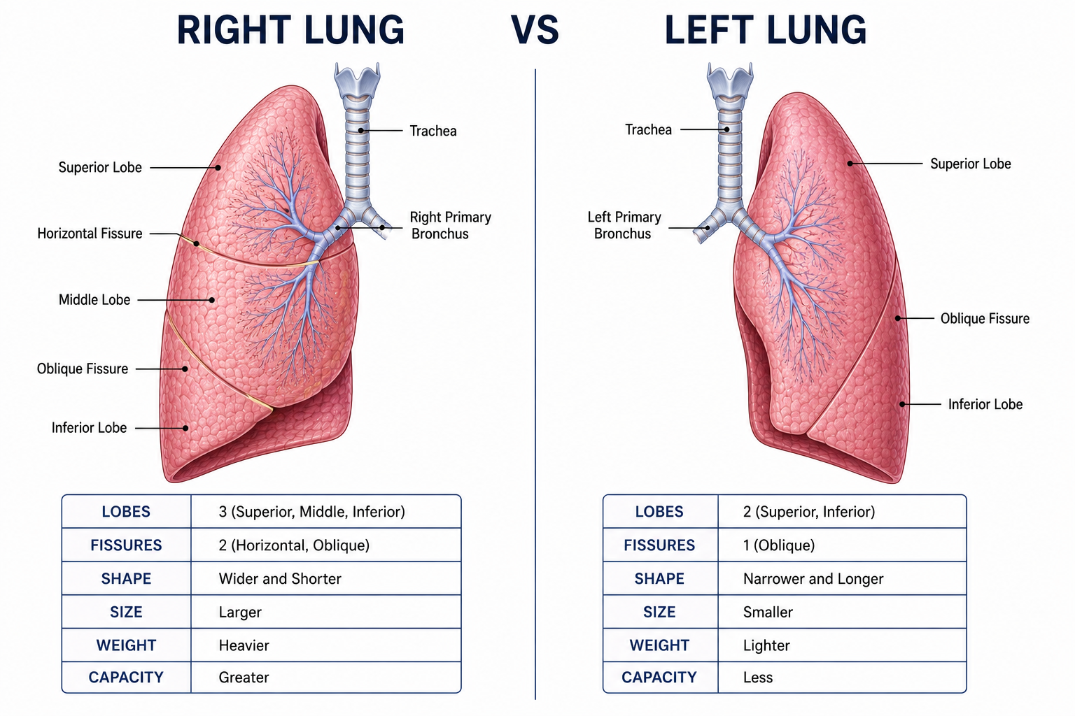
Right Lung
- ✅ 3 Lobes
- 📏 Larger
- ↔️ Wider
- ⬇️ Shorter
- 🚫 No Cardiac Notch
Left Lung
- ✅ 2 Lobes
- 📏 Smaller
- ↔️ Narrower
- ⬆️ Longer
- 💓 Cardiac Notch Present
🫁 Lobes of the Lungs
The lungs are divided into sections called lobes. These lobes organize the lungs into separate regions, allowing air to be distributed efficiently and enabling one part of the lung to function even if another becomes damaged. The right lung contains three lobes, while the left lung has two lobes because it shares space with the heart.
Right Lung
The upper lobe of the right lung. It receives fresh air and participates in normal gas exchange.
The smallest lobe of the right lung, located between the superior and inferior lobes. It helps distribute air throughout the lung.
The largest lobe of the right lung. It lies near the diaphragm and performs efficient gas exchange.
Left Lung
The upper lobe of the left lung. It contains the cardiac notch, which creates space for the heart.
The lower lobe of the left lung. It occupies much of the lower portion of the lung and carries out gas exchange like all other lobes.
💡 Did You Know?
Although the lungs are divided into different lobes, every lobe performs the same essential function—exchanging oxygen and carbon dioxide. The lobes mainly help organize the lungs and allow different regions to work independently. This means that if one lobe becomes infected or damaged, the remaining lobes can often continue functioning normally.
🫁 Pleura
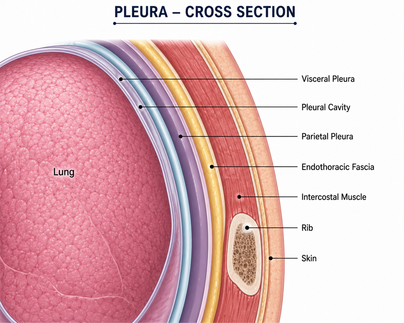
The pleura is a double-layered membrane that surrounds each lung and lines the inside of the chest cavity. Between these two layers is a very thin space called the pleural cavity, which contains a small amount of pleural fluid. Together, these structures protect the lungs and allow them to move smoothly during breathing without rubbing against the chest wall.
Visceral Pleura
The visceral pleura is the inner layer of the pleura. It is tightly attached to the surface of the lungs and moves with them during every breath.
Pleural Cavity
The pleural cavity is the narrow space between the visceral and parietal pleura. It contains a very small amount of pleural fluid.
Pleural Fluid
Pleural fluid acts as a natural lubricant. It reduces friction and allows the lungs to glide smoothly against the chest wall during inhalation and exhalation.
Parietal Pleura
The parietal pleura is the outer layer of the pleura. It lines the inside of the rib cage, diaphragm, and chest cavity, protecting the lungs and supporting their movement.
🌬 Why is the Pleura Important?
During breathing, the lungs continuously expand and contract. Without the pleura, the lungs would rub directly against the ribs, causing pain and damage. The pleural fluid between the two pleural layers acts like a lubricant, allowing the lungs to move effortlessly while maintaining a gentle suction that keeps them attached to the chest wall.
🌳 Inside the Lungs
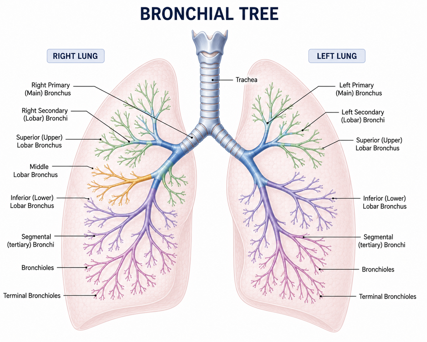
Inside the lungs, the airways branch repeatedly like an upside-down tree. Each branch becomes progressively smaller, allowing air to reach millions of tiny alveoli, where oxygen enters the blood and carbon dioxide leaves the body. This branching pattern is called the bronchial tree.
Lungs
The paired respiratory organs that contain the complete bronchial tree and millions of alveoli.
Primary Bronchi
The trachea divides into the right and left primary bronchi, one entering each lung.
Secondary Bronchi
Each primary bronchus divides into secondary bronchi, supplying individual lobes of the lungs.
Bronchioles
Thousands of tiny bronchioles distribute air throughout every region of the lungs.
Alveolar Ducts
Small passages that connect bronchioles to clusters of alveolar sacs.
Alveolar Sacs
Tiny clusters containing numerous alveoli, greatly increasing the surface area for gas exchange.
Alveoli
Microscopic air sacs where oxygen diffuses into the blood and carbon dioxide diffuses out of the blood.
🫧 Alveoli Explorer
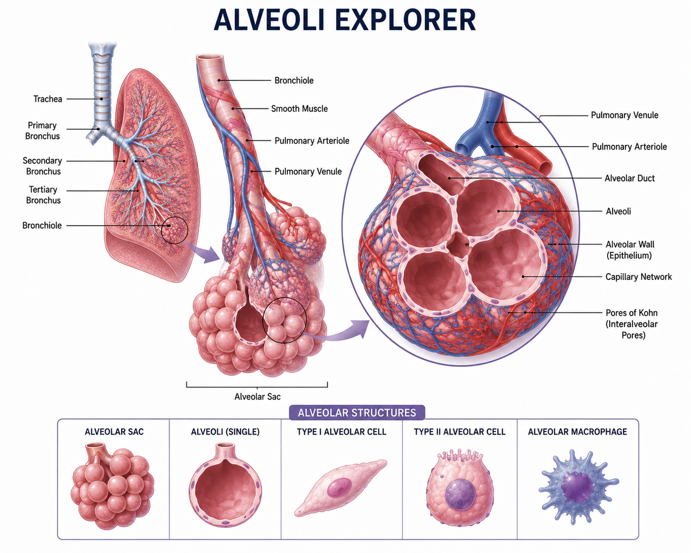
The alveoli are tiny microscopic air sacs located at the ends of the bronchioles. They are the functional units of the lungs where gas exchange occurs. Humans have approximately 300 million alveoli, providing an enormous surface area for oxygen and carbon dioxide to diffuse efficiently.
Thin Walls
Each alveolus has walls only one cell thick, allowing oxygen and carbon dioxide to diffuse rapidly.
Moist Lining
A thin layer of moisture covers the alveoli, helping oxygen dissolve before passing into the bloodstream.
Capillary Network
Every alveolus is wrapped in tiny blood capillaries, bringing deoxygenated blood close to the air inside the lungs.
Large Surface Area
Together, millions of alveoli create approximately 70 m² of gas-exchange surface—about the size of half a tennis court.
🌬 Gas Exchange in Action
Oxygen moves from the air inside the alveolus into the blood, while carbon dioxide moves from the blood into the alveolus before being exhaled.
💨 Lung Volumes & Capacities
During breathing, the lungs hold different amounts of air depending on the activity being performed. These measurements are known as lung volumes and lung capacities, and they help doctors evaluate how well the lungs are functioning.
Tidal Volume (TV)
The amount of air inhaled or exhaled during a normal, relaxed breath.
Inspiratory Reserve Volume (IRV)
The extra amount of air that can be inhaled after taking a normal breath.
Expiratory Reserve Volume (ERV)
The extra amount of air that can be forcefully exhaled after a normal breath.
Residual Volume (RV)
The air that always remains inside the lungs, even after maximum exhalation.
Vital Capacity (VC)
The greatest amount of air a person can exhale after taking the deepest breath possible.
Total Lung Capacity (TLC)
The maximum amount of air the lungs can hold after a full inhalation.
❤️ Pulmonary Circulation
Pulmonary circulation is the pathway through which deoxygenated blood travels from the heart to the lungs, becomes oxygenated, and then returns to the heart as oxygenated blood. This process ensures that fresh oxygen is supplied to the body while carbon dioxide is removed.
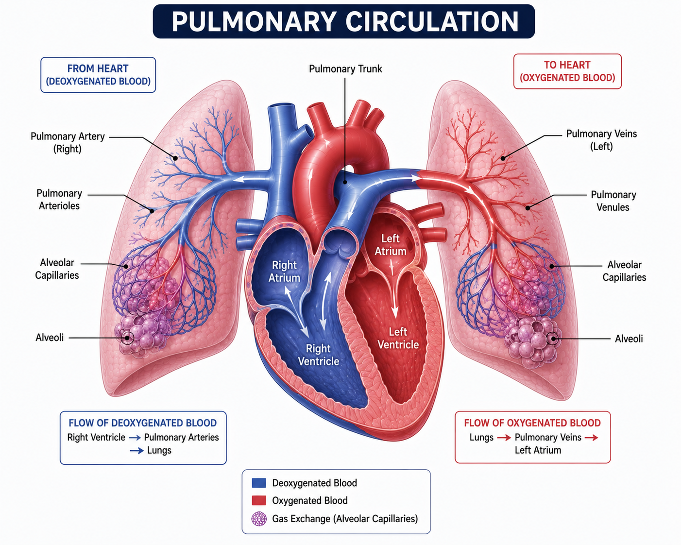
Heart
The right side of the heart pumps deoxygenated blood toward the lungs.
Pulmonary Artery
Carries deoxygenated blood from the heart to the lungs.
Lungs
Gas exchange occurs in the alveoli. Oxygen enters the blood while carbon dioxide leaves it.
Pulmonary Veins
Carry oxygenated blood back to the heart.
Heart
The left side of the heart receives oxygenated blood and pumps it to the rest of the body.
🔵 Blue: Deoxygenated Blood
🔴 Red: Oxygenated Blood
🫁 How the Lungs Expand
Breathing is made possible by the coordinated action of the diaphragm, intercostal muscles, and rib cage. These structures change the size of the chest cavity, causing the lungs to either expand during inhalation or shrink during exhalation.
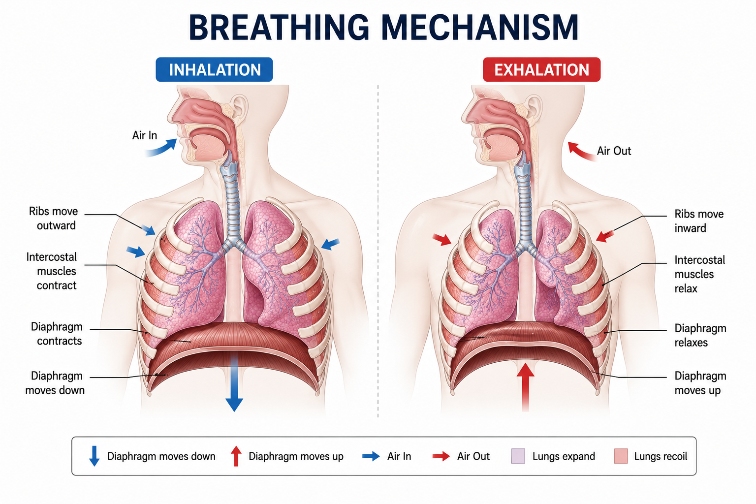
🎚 Breathing Simulator
🫁 Neutral
Move the slider to visualize the transition between Inhalation and Exhalation.
Inhalation
- ⬇️ Diaphragm contracts and moves downward
- ⬆️ Rib cage moves upward and outward
- 🫁 Lungs expand
- 🌬 Air flows into the lungs
Exhalation
- ⬆️ Diaphragm relaxes and moves upward
- ⬇️ Rib cage moves downward and inward
- 🫁 Lungs recoil
- 💨 Air leaves the lungs
💚 Keeping Your Lungs Healthy
Healthy lungs are essential for supplying oxygen to every cell in the body. Simple daily habits can strengthen the respiratory system, improve breathing, and reduce the risk of lung diseases.
Avoid Smoking
Smoking damages lung tissue, destroys alveoli, and greatly increases the risk of chronic lung diseases and lung cancer.
Exercise Regularly
Physical activity strengthens the lungs, improves breathing efficiency, and helps the body use oxygen more effectively.
Practice Deep Breathing
Deep breathing exercises expand the lungs, improve airflow, and help maintain healthy lung function.
Reduce Exposure to Polluted Air
Avoid smoke, dust, and harmful chemicals whenever possible, and wear a mask in heavily polluted environments.
Eat a Balanced Diet
Fruits, vegetables, whole grains, and foods rich in vitamins help keep the lungs and immune system healthy.
Stay Hydrated
Drinking enough water keeps mucus thin, making it easier for the lungs to remove dust and germs.
Stay Up to Date with Vaccinations
Recommended vaccines help protect against infections such as influenza and pneumonia that can seriously affect the lungs.
🩺 Clinical Connections
Understanding how the lungs work helps explain many everyday experiences and common medical conditions. These real-life connections show why keeping the lungs healthy is so important.
Why do lungs float?
Healthy lungs contain millions of tiny air-filled alveoli, making them less dense than water.
Why is the left lung smaller?
The heart occupies space on the left side of the chest, so the left lung has only two lobes and a cardiac notch.
Why is breathing harder at high altitude?
High altitudes contain less oxygen, making it more difficult for the body to obtain enough oxygen with each breath.
Why does exercise improve lung efficiency?
Regular physical activity strengthens the respiratory muscles and improves the body's ability to use oxygen.
Why do doctors listen to your lungs?
A stethoscope helps detect abnormal breathing sounds that may indicate infections, asthma, or fluid in the lungs.
Why does asthma cause wheezing?
Inflamed and narrowed airways make it difficult for air to pass, producing a whistling sound called wheezing.
Why is pneumonia dangerous?
Pneumonia fills the alveoli with fluid or pus, reducing oxygen exchange and making breathing difficult.
Why is smoking harmful?
Smoking damages the airways and alveoli, reducing lung function and greatly increasing the risk of lung disease.
Why can one lung keep you alive?
One healthy lung can usually provide enough oxygen for everyday life, although physical performance may be reduced.
Why is oxygen therapy used?
Oxygen therapy increases the amount of oxygen reaching the blood when the lungs cannot supply enough on their own.
📊 Amazing Lung Statistics
The lungs work continuously throughout life, performing millions of tiny tasks every day to keep the body supplied with oxygen. These fascinating facts reveal just how incredible your respiratory system really is.
Two Lungs
Humans normally have two lungs working together to oxygenate blood.
Alveoli
Around 300 million alveoli provide an enormous surface for gas exchange.
Gas Exchange Area
The lungs have a surface area roughly the size of a tennis court.
Breaths Every Day
An average person breathes approximately twenty thousand times daily.
Right Lung Lobes
The right lung consists of three separate lobes.
Left Lung Lobes
The left lung has only two lobes because the heart occupies space.
Cardiac Output
Every drop of blood pumped by the heart passes through the lungs.
Normal Breathing Rate
Healthy adults usually breathe between 12 and 20 times each minute.
Amazing Liver Facts
The liver is the largest internal organ and one of the busiest organs in the human body. It performs hundreds of essential functions every single day!
The liver performs over 500 different functions to keep your body healthy.
It is the largest internal organ in the human body.
An adult human liver weighs about 1.4–1.8 kg.
The liver receives blood from two different blood vessels—the hepatic artery and the portal vein.
The liver has an incredible ability to regenerate itself after injury or surgery.
The liver continuously produces bile, which helps digest fats.
Your liver produces around 600–1000 mL of bile every day.
It stores extra glucose as glycogen and releases it when your body needs energy.
The liver removes harmful toxins from the blood before they can damage the body.
Most medicines are broken down by the liver before leaving the body.
The liver contains special immune cells called Kupffer cells that destroy harmful bacteria.
Almost 13% of your body's blood can be found in the liver at any moment.
The liver produces many important blood proteins, including albumin and clotting factors.
Without the liver, your blood would have difficulty clotting after an injury.
The liver helps maintain a stable body temperature because many chemical reactions occur inside it.
It converts excess amino acids into useful substances and safely removes their waste products.
The liver plays a major role in cholesterol production and regulation.
Before birth, the liver helps produce red blood cells in the developing baby.
The liver is located mainly on the right side of the abdomen, just beneath the diaphragm.
The liver is the only major internal organ capable of regenerating a large portion of itself after damage.
💡 Did you know?
Scientists have discovered that the human liver can regrow to nearly its original size even if up to 70% of it is surgically removed, making it the only major internal organ with such an extraordinary regenerative ability.