Interactive Heart Labelled Illustration
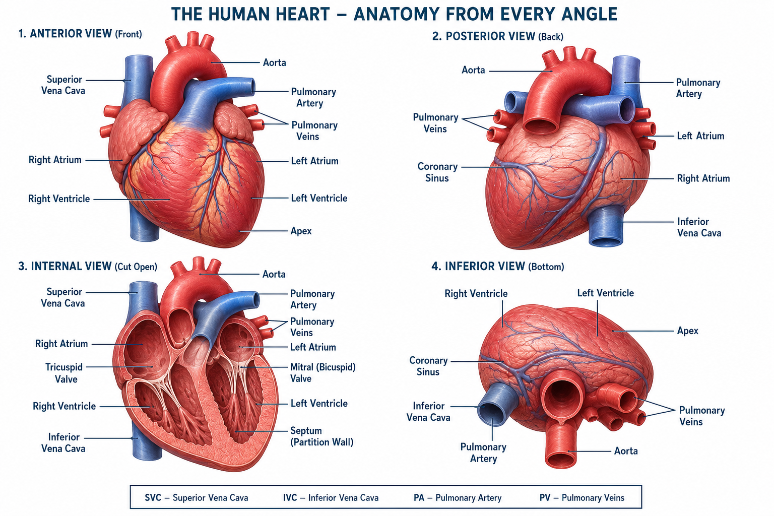
Functions of the Heart
The heart is the central pumping organ of the circulatory system. It continuously pumps blood throughout the body, ensuring that every cell receives oxygen and nutrients while removing waste products. Besides pumping blood, the heart also plays a vital role in maintaining homeostasis.
❤️ Pumps Oxygenated Blood
The left side of the heart pumps oxygen-rich blood received from the lungs to every organ and tissue through the aorta, ensuring that body cells receive the oxygen needed for cellular respiration.
🩸 Pumps Deoxygenated Blood
The right side of the heart pumps oxygen-poor blood to the lungs through the pulmonary artery, where carbon dioxide is removed and fresh oxygen is absorbed.
🫁 Maintains Blood Circulation
By continuously contracting and relaxing, the heart keeps blood circulating through arteries, veins, and capillaries, supplying every cell of the body with essential substances.
🍎 Delivers Nutrients
Blood pumped by the heart carries nutrients absorbed from the digestive system to body tissues, providing the energy and raw materials required for growth and repair.
🧪 Removes Waste Products
The heart transports blood carrying carbon dioxide and metabolic wastes to organs such as the lungs, kidneys, and liver, where these wastes are removed from the body.
💉 Transports Hormones
Hormones released by endocrine glands are carried by the bloodstream, allowing the heart to distribute these important chemical messengers throughout the body.
🛡 Supports Immunity
The heart circulates white blood cells and antibodies throughout the body, helping the immune system detect and destroy harmful microorganisms.
🌡 Regulates Body Temperature
Blood circulation helps distribute heat evenly throughout the body, maintaining a stable body temperature despite changes in the external environment.
⚖ Maintains Blood Pressure
The pumping action of the heart generates the blood pressure required to move blood efficiently through blood vessels, ensuring adequate circulation to all body organs.
💓 Sustains Life
Every heartbeat supplies oxygen and nutrients necessary for survival. Without the heart's continuous pumping action, body cells would quickly die due to a lack of oxygen.
External Anatomy of the Heart
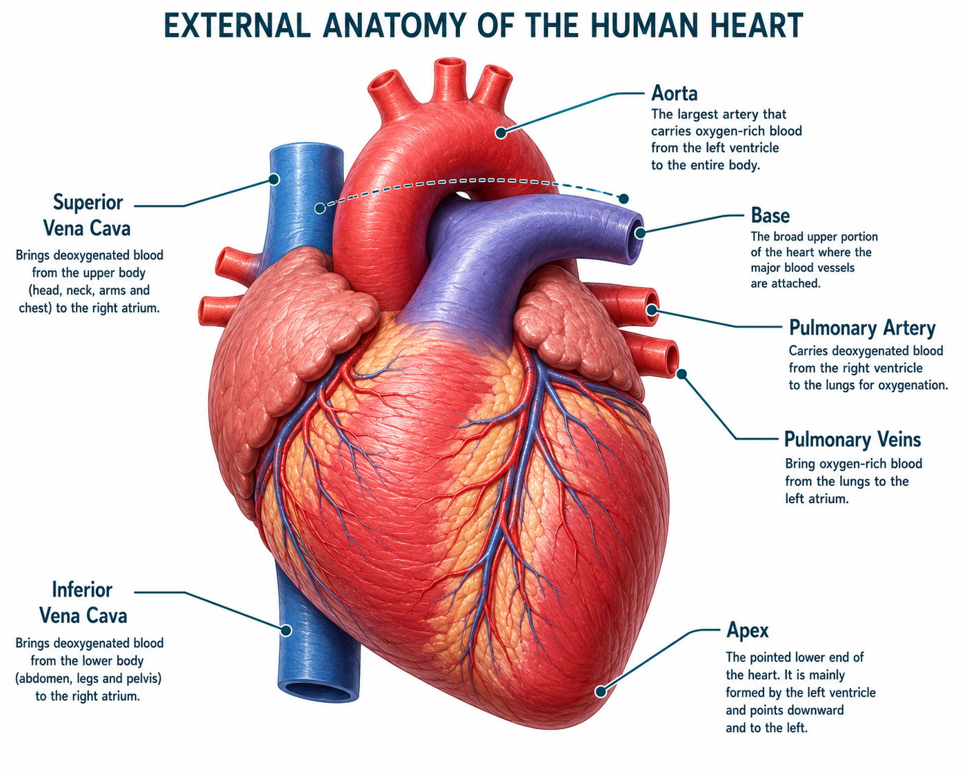
The external anatomy of the heart includes the major blood vessels and visible structures responsible for transporting blood between the heart, lungs, and the rest of the body. Understanding these structures helps explain how blood enters and leaves the heart during circulation.
🫀 Aorta
The aorta is the largest artery in the body. It carries oxygen-rich blood from the left ventricle to all parts of the body.
🔵 Superior Vena Cava
The superior vena cava returns deoxygenated blood from the head, neck, arms, and upper body to the right atrium.
🔵 Inferior Vena Cava
The inferior vena cava carries oxygen-poor blood from the lower body back to the right atrium.
🫁 Pulmonary Artery
The pulmonary artery transports deoxygenated blood from the right ventricle to the lungs for oxygenation.
❤️ Pulmonary Veins
The pulmonary veins return oxygen-rich blood from the lungs to the left atrium.
📍 Apex
The apex is the pointed lower tip of the heart. It is mainly formed by the left ventricle and points downward and to the left.
🏛 Base
The base is the broad upper portion of the heart where the major blood vessels are attached.
Internal Anatomy of the Heart
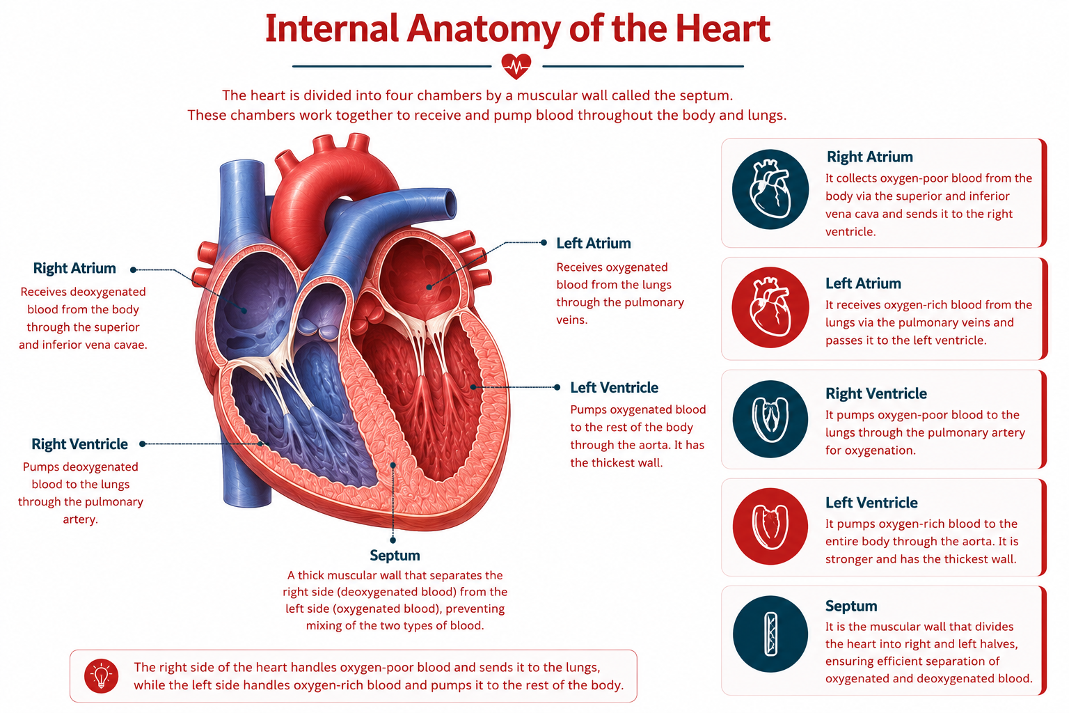
The internal anatomy of the heart consists of four chambers separated by a muscular wall called the septum. These chambers work together to receive, pump, and circulate blood efficiently throughout the body.
🔵 Right Atrium
The right atrium is the upper right chamber of the heart. It receives deoxygenated blood from the body through the superior and inferior vena cava before passing it to the right ventricle.
🔴 Left Atrium
The left atrium is the upper left chamber. It receives oxygen-rich blood from the lungs through the pulmonary veins and sends it to the left ventricle.
🔵 Right Ventricle
The right ventricle pumps deoxygenated blood to the lungs through the pulmonary artery, where the blood becomes oxygenated.
🔴 Left Ventricle
The left ventricle is the largest and strongest chamber of the heart. It pumps oxygenated blood into the aorta, supplying the entire body.
🧱 Septum
The septum is the thick muscular wall that separates the right and left sides of the heart. It prevents oxygen-rich and oxygen-poor blood from mixing.
Heart Valves
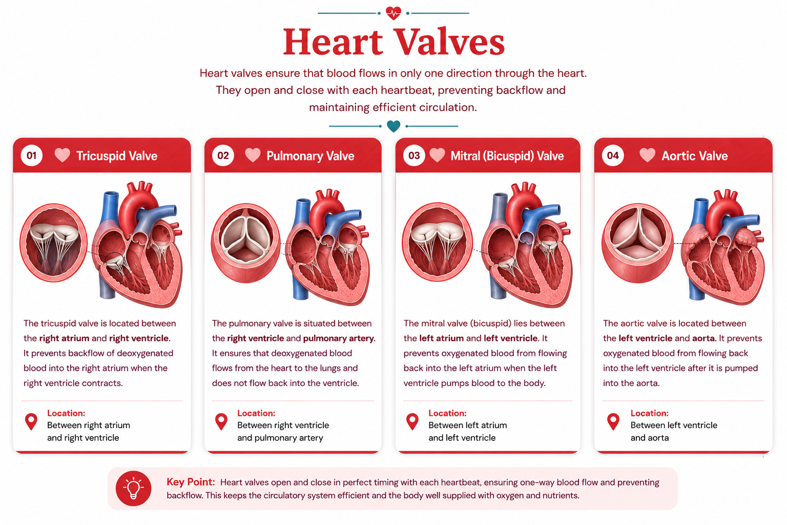
The heart valves act like one-way doors that ensure blood flows in the correct direction. They open to allow blood to pass through and close immediately afterward to prevent backflow.
Tricuspid Valve
Located between the right atrium and the right ventricle, the tricuspid valve prevents blood from flowing backward into the right atrium during ventricular contraction.
Pulmonary Valve
Situated between the right ventricle and the pulmonary artery, it ensures blood flows only toward the lungs and prevents its return to the heart.
Mitral (Bicuspid) Valve
Found between the left atrium and the left ventricle, this valve prevents oxygen-rich blood from flowing backward into the left atrium.
Aortic Valve
Located between the left ventricle and the aorta, the aortic valve prevents blood from flowing back into the left ventricle after it has entered the aorta.
Blood Flow Through the Heart
Blood follows a continuous one-way pathway through the heart, lungs, and body. The heart valves ensure that blood always moves in the correct direction, preventing backflow.
Body
Deoxygenated blood returns from body tissues.
Superior & Inferior Vena Cava
Carry oxygen-poor blood into the heart.
Right Atrium
Receives deoxygenated blood.
Tricuspid Valve
Allows blood to enter the right ventricle.
Right Ventricle
Pumps blood toward the lungs.
Pulmonary Valve
Prevents blood from flowing backward.
Pulmonary Artery
Carries blood to the lungs.
Lungs
Blood receives oxygen and releases carbon dioxide.
Pulmonary Veins
Carry oxygen-rich blood back to the heart.
Left Atrium
Receives oxygenated blood.
Mitral Valve
Directs blood into the left ventricle.
Left Ventricle
The strongest chamber pumps blood to the body.
Aortic Valve
Prevents blood from returning to the ventricle.
Aorta
The largest artery distributes oxygen-rich blood.
Body
Cells receive oxygen and nutrients, and the cycle repeats.
Layers of the Heart Wall
The heart wall is made up of three distinct layers. Each layer performs a specialized function that protects the heart, enables powerful contractions, and ensures smooth blood flow throughout the circulatory system.
Endocardium
The endocardium is the innermost layer of the heart. It forms a smooth lining for the heart chambers and valves, reducing friction as blood flows through the heart.
Myocardium
The myocardium is the middle and thickest layer of the heart wall. It consists of cardiac muscle tissue responsible for the powerful contractions that pump blood throughout the body.
Epicardium
The epicardium is the outermost protective layer of the heart. It forms part of the pericardium and helps protect the heart while reducing friction during each heartbeat.
Cardiac Cycle
The cardiac cycle is the sequence of events that occurs during a single heartbeat. It consists of three main phases that ensure blood is pumped efficiently through the heart and the rest of the body.
Diastole
During diastole, the heart muscles relax. All four chambers fill with blood while the atrioventricular valves remain open.
Atrial Systole
During atrial systole, the atria contract, pushing the remaining blood into the ventricles before ventricular contraction begins.
Ventricular Systole
During ventricular systole, the ventricles contract forcefully, pumping blood into the pulmonary artery and the aorta.
Cardiac Conduction System
The cardiac conduction system is a specialized network of electrical tissues that generates and conducts electrical impulses. These impulses coordinate the heartbeat, ensuring that the atria and ventricles contract in the correct sequence.
SA Node
The Sinoatrial (SA) Node is the heart's natural pacemaker. It generates electrical impulses that initiate each heartbeat.
AV Node
The Atrioventricular (AV) Node receives impulses from the SA node and briefly delays them, allowing the ventricles to fill with blood before contracting.
Bundle of His
The Bundle of His carries electrical signals from the AV node through the septum toward the ventricles.
Purkinje Fibers
Purkinje Fibers rapidly distribute electrical impulses throughout the ventricles, causing a strong and coordinated ventricular contraction.
Coronary Circulation
The coronary circulation is the network of blood vessels that supplies the heart muscle (myocardium) with oxygen and nutrients while removing carbon dioxide and metabolic wastes. Without this dedicated blood supply, the heart would be unable to function effectively.
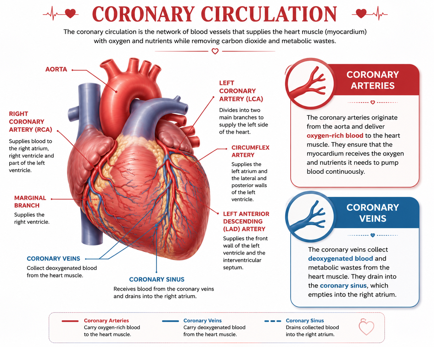
Coronary Arteries
The coronary arteries branch from the aorta and deliver oxygen-rich blood to the heart muscle. They nourish the myocardium with the oxygen and nutrients required for continuous contraction.
Coronary Veins
The coronary veins collect deoxygenated blood and metabolic wastes from the heart muscle. They empty into the coronary sinus, which drains into the right atrium.
Cardiac Output
Cardiac Output (CO) is the amount of blood pumped by the heart in one minute. It depends on two important factors: Heart Rate (HR) and Stroke Volume (SV).
🫀 Cardiac Output Formula
Heart Rate (HR): Number of heartbeats per minute.
Stroke Volume (SV): Amount of blood pumped by one ventricle during each heartbeat.
📘 Example Calculation
Heart Rate: 72 beats/minute
Stroke Volume: 70 mL/beat
CO = 72 × 70
≈ 5 Litres of blood per minute
💡 An average healthy adult heart pumps approximately 5 litres of blood every minute, and this amount increases during exercise.
Heart Sounds
During every heartbeat, the heart produces two characteristic sounds: "Lub" and "Dub". These sounds are caused by the closing of the heart valves, not by the heart muscle itself.
Lub (First Heart Sound)
The "Lub" sound is produced when the tricuspid valve and mitral (bicuspid) valve close at the beginning of ventricular systole.
Dub (Second Heart Sound)
The "Dub" sound is produced when the aortic valve and pulmonary valve close at the beginning of ventricular diastole.
💡 Did You Know?
Doctors use a stethoscope to listen to the Lub-Dub sounds. Any extra or abnormal sounds are called heart murmurs, which may indicate problems with the heart valves or blood flow.
Amazing Heart Facts
Here are some fascinating facts about the human heart!
Your heart beats about 100,000 times every day.
The heart pumps nearly 7,500 litres (2,000 gallons) of blood through your body every day.
Your heart has its own electrical conduction system, allowing it to beat without instructions from the brain.
The left ventricle is the strongest chamber because it pumps blood to the entire body.
Your heart is approximately the same size as your clenched fist.
A newborn baby's heart beats much faster than an adult's, often reaching 120–160 beats per minute.
During intense exercise, your heart can pump up to 35 litres of blood every minute.
The right side of the heart pumps oxygen-poor blood to the lungs.
The left side of the heart pumps oxygen-rich blood to every part of your body.
The familiar "Lub-Dub" sound is produced by the closing of the heart valves.
An average adult heart weighs only about 250–350 grams.
Your heart rate naturally slows down while you sleep because your body requires less oxygen.
Laughter improves blood circulation and can temporarily increase your heart rate in a healthy way.
The human heart starts beating only about 22 days after fertilization, long before birth.
The heart receives its own blood supply through the coronary arteries.
Doctors use a stethoscope to listen for heart sounds and detect possible heart disorders.
If all the blood vessels in your body were joined together, they would stretch for about 100,000 kilometres.
The blue whale has the largest heart of any animal, weighing more than 180 kilograms.
Your heart never truly rests; it continues working every second throughout your entire life.
Your heart generates enough pressure to squirt blood nearly 9 metres (30 feet) if blood escaped from a major artery.
💡 Did you know?
During an average lifetime, your heart beats over 3 billion times and pumps enough blood to fill about three Olympic-sized swimming pools—all without ever taking a single break.