Interactive Carnial Nerves present in the brain Labelled Illustration
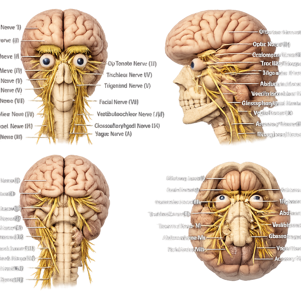
The cranial nerves are 12 pairs of peripheral nerves that emerge directly from the brain and brainstem. Unlike the spinal nerves, which arise from the spinal cord, cranial nerves bypass the spinal cord and connect the brain directly to various parts of the head, neck, and some organs in the chest and abdomen. They carry sensory, motor, and autonomic signals, allowing the brain to receive information from the body's sense organs and control the movement of muscles and glands. These nerves are responsible for essential functions such as vision, hearing, smell, taste, eye movements, facial expressions, swallowing, speech, and the regulation of several involuntary body functions. Each cranial nerve is identified by a Roman numeral (I–XII) according to its position and has a specific structure and function. Together, the cranial nerves play a vital role in communication between the brain and different parts of the body, ensuring the proper functioning of many sensory and motor activities.
The 12 pairs of cranial nerves arise directly from the brain and brainstem. Each nerve has a specific function, such as carrying sensory information, controlling muscle movements, or performing both sensory and motor functions. They are numbered using Roman numerals (I–XII) according to their position from the front to the back of the brain.
Type: Sensory
The olfactory nerve carries smell sensations from the
nose to the brain. It allows us to detect and recognize different
odors.
Type: Sensory
The optic nerve transmits visual information from the
retina to the brain, making vision possible.
Type: Motor
Controls most eye movements, raises the upper eyelid, and regulates
pupil constriction and lens focusing.
Type: Motor
Controls the superior oblique muscle, allowing the eye to move
downward and inward.
Type: Mixed
Provides sensation to the face and controls the muscles used for
chewing.
Type: Motor
Controls the lateral rectus muscle, allowing the eye to move
outward.
Type: Mixed
The facial nerve controls the muscles of facial expression, carries the sense of taste from the front two-thirds of the tongue, and helps regulate tear and saliva production.
Type: Sensory
The vestibulocochlear nerve is responsible for hearing and balance, carrying sound and equilibrium information from the inner ear to the brain.
Type: Mixed
The glossopharyngeal nerve provides taste from the back one-third of the tongue, assists in swallowing, and helps regulate salivary gland function.
Type: Mixed
The vagus nerve controls many automatic body functions, including the heart, lungs, digestive system, and also helps with speech and swallowing.
Type: Motor
The accessory nerve controls the sternocleidomastoid and trapezius muscles, allowing movements of the head, neck, and shoulders.
Type: Motor
The hypoglossal nerve controls the muscles of the tongue, making it essential for speech, chewing, and swallowing.
"Oh, Oh, Oh, To Touch And Feel Very Good Velvet, Such Heaven"
"Old Owls Often Try Teaching Angry Foxes Very Good Vocabulary Skills Happily"
"Our Outstanding Office Teacher Always Finds Very Good Volunteers Serving Happily."
The 12 cranial nerves are classified according to the type of information they carry. Some carry only sensory impulses, some control motor functions, while others perform both sensory and motor functions.
These nerves carry sensory information from the sensory organs to the brain.
These nerves control the movement of muscles in the eyes, tongue, neck, and shoulders.
These nerves perform both sensory and motor functions.
"Some Say Marry Money, But My Brother Says Big Brains Matter More."
Some → CN I (Sensory)
Say → CN II (Sensory)
Marry → CN III (Motor)
Money → CN IV (Motor)
But → CN V (Both)
My → CN VI (Motor)
Brother → CN VII (Both)
Says → CN VIII (Sensory)
Big → CN IX (Both)
Brains → CN X (Both)
Matter → CN XI (Motor)
More → CN XII (Motor)
S = Sensory | M = Motor | B = Both (Mixed)
Here are some fascinating facts about the Cranial Nerves!
The olfactory nerve (CN I) is the only cranial nerve directly exposed to the external environment.
The optic nerve (CN II) is actually an extension of the brain, not a typical peripheral nerve.
Humans have 12 pairs of cranial nerves, all of which arise directly from the brain or brainstem.
The vagus nerve (CN X) is the longest cranial nerve, extending from the brain to the chest and abdomen.
The facial nerve (CN VII) controls most of the muscles responsible for facial expressions.
Taste is carried by three different cranial nerves: the Facial (CN VII), Glossopharyngeal (CN IX), and Vagus (CN X) nerves.
The vestibulocochlear nerve (CN VIII) helps maintain both hearing and balance.
Three cranial nerves—CN III, CN IV, and CN VI—work together to control eye movements.
The accessory nerve (CN XI) allows you to shrug your shoulders and turn your head.
The hypoglossal nerve (CN XII) controls tongue movements needed for speaking, chewing, and swallowing.
Cranial nerves transmit electrical impulses between the brain and different parts of the head, neck, and body.
The vestibulocochlear nerve contains separate branches for hearing (cochlear) and balance (vestibular).
The trigeminal nerve (CN V) is the largest cranial nerve and provides sensation to most of the face.
Dentists often numb branches of the trigeminal nerve during dental procedures.
The vagus nerve helps slow the heart rate and supports normal digestion.
The glossopharyngeal and vagus nerves work together during swallowing.
Not all cranial nerves are the same—some are sensory, some are motor, and others are mixed.
Medical students often memorize the cranial nerves using the famous mnemonic: "Oh, Oh, Oh, To Touch And Feel Very Good Velvet, Such Heaven."
The vagus nerve is called the "wandering nerve" because it travels farther than any other cranial nerve.
Doctors routinely examine the cranial nerves during neurological examinations to assess brain and nervous system function.
The vagus nerve (CN X) is often called the "wandering nerve" because it travels farther than any other cranial nerve. It extends from the brain all the way to the heart, lungs, stomach, liver, intestines, and many other organs, helping regulate breathing, heartbeat, digestion, and other vital body functions.
The olfactory nerve (CN I) is the first cranial nerve and is a sensory nerve. It is responsible for the sense of smell, carrying odor signals from the nose to the brain. It allows us to detect and identify different smells in our surroundings.
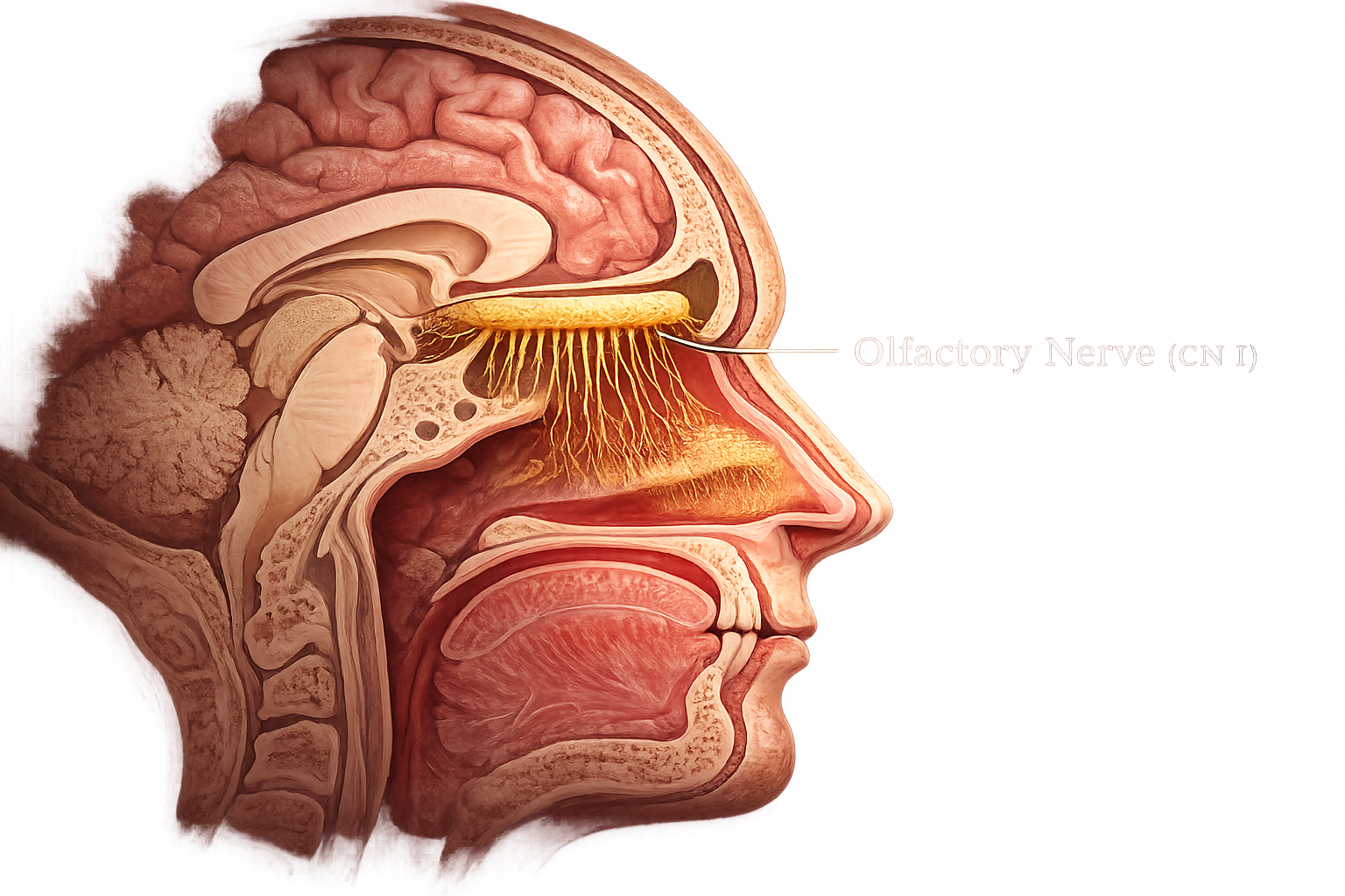
Olfactory receptors are specialized sensory cells located in the olfactory epithelium inside the roof of the nasal cavity. They detect odor molecules from the air and convert them into electrical nerve impulses, initiating the sense of smell.
The tiny olfactory nerve fibers (fila olfactoria) are bundles of sensory axons that pass through the cribriform plate of the ethmoid bone. They carry smell signals from the olfactory receptors to the olfactory bulb.
The olfactory bulb is the first processing center for smell. It receives nerve impulses from the olfactory nerve fibers, processes the information, and sends it toward higher brain regions for odor recognition.
The olfactory tract is a bundle of nerve fibers extending from the olfactory bulb to the brain. It carries processed smell information to the olfactory cortex and other brain regions involved in memory and emotion.
The olfactory nerve (CN I) is an important sensory nerve that provides the sense of smell. It enables the brain to recognize different odors, enhances the perception of flavor, and helps detect potentially dangerous substances in the environment.
The optic nerve (CN II) is the second cranial nerve and is a sensory nerve. It is responsible for the sense of vision, carrying visual information from the retina of the eye to the brain. It enables us to see objects, recognize colors, and perceive light.
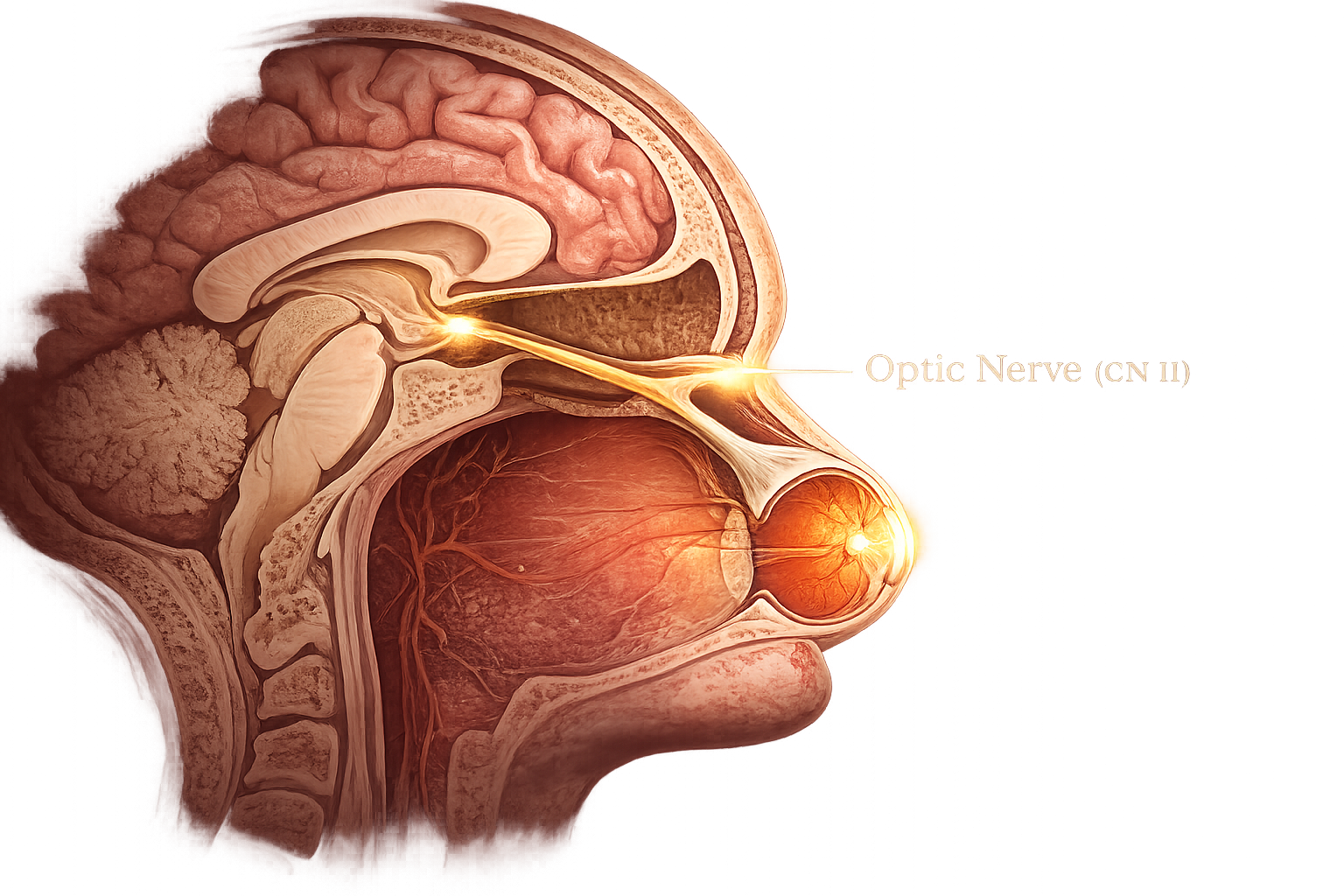
The retina is the light-sensitive layer located at the back of the eye. It contains rods and cones, which detect light and convert it into electrical nerve impulses. These impulses are the first step in the process of vision.
The optic disc, also called the blind spot, is the point where the optic nerve leaves the retina. It contains no photoreceptors, so light falling on this area cannot be detected.
The optic nerve (CN II) is formed by more than one million nerve fibers. It carries visual impulses from the retina to the brain, making it the main pathway for the sense of vision.
The optic chiasm is an X-shaped structure where some optic nerve fibers cross to the opposite side of the brain. This crossing allows information from both eyes to be combined, helping produce binocular vision.
The optic tract carries visual information from the optic chiasm to the brain. It transmits signals primarily to the lateral geniculate nucleus (LGN) of the thalamus for further processing.
The visual cortex, located in the occipital lobe of the brain, is the final destination of visual impulses. Here, the brain interprets information about color, shape, movement, and depth, allowing us to see and understand our surroundings.
The optic nerve (CN II) is a vital sensory nerve that provides the sense of vision. It carries visual information from the eyes to the brain, allowing us to see, recognize objects, and respond to changes in light.
The oculomotor nerve (CN III) is the third cranial nerve and is primarily a motor nerve. It controls most of the eye movements, raises the upper eyelid, and helps adjust the pupil size and lens shape for clear vision.
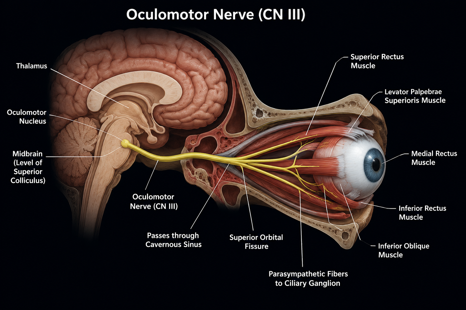
The oculomotor nucleus is located in the midbrain at the level of the superior colliculus. It contains motor neurons that give rise to the oculomotor nerve and control most of the eye muscles responsible for eye movement.
The Edinger–Westphal nucleus is the parasympathetic nucleus of the oculomotor nerve. It sends nerve fibers that control pupil constriction and the accommodation of the lens for focusing on nearby objects.
The oculomotor nerve emerges from the front of the midbrain and travels through the cavernous sinus before entering the orbit. It carries both motor and parasympathetic fibers to the eye.
The superior orbital fissure is an opening in the skull through which the oculomotor nerve enters the orbit. It allows the nerve to reach the muscles that control eye movement and the upper eyelid.
The oculomotor nerve supplies the superior rectus, inferior rectus, medial rectus, inferior oblique, and the levator palpebrae superioris. These muscles move the eyeball in different directions and raise the upper eyelid.
The ciliary ganglion receives parasympathetic fibers from the oculomotor nerve and sends signals to the sphincter pupillae and ciliary muscles. These muscles constrict the pupil and adjust the lens for clear vision at different distances.
The oculomotor nerve (CN III) is an important motor nerve that controls most eye movements, raises the upper eyelid, and regulates the pupil and lens. It plays a vital role in maintaining clear vision and proper eye coordination.
The trochlear nerve (CN IV) is the fourth cranial nerve and is a motor nerve. It controls the superior oblique muscle of the eye, allowing the eye to move downward and inward. It plays an important role in coordinated eye movements and stable vision.
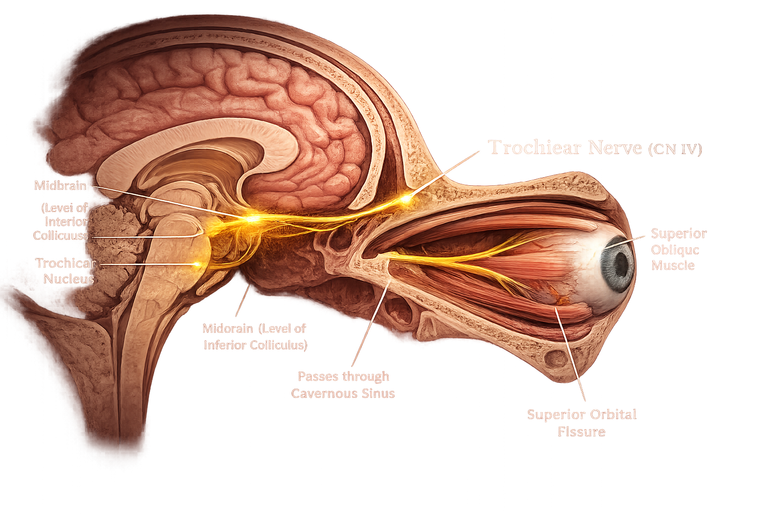
The trochlear nucleus is located in the midbrain, just below the oculomotor nucleus. It contains motor neurons that give rise to the trochlear nerve and initiate the nerve impulses responsible for controlling eye movement.
The trochlear nerve (CN IV) is the smallest cranial nerve and the only cranial nerve that emerges from the back (dorsal surface) of the brainstem. It carries motor fibers to a single eye muscle.
Before leaving the brainstem, the fibers of the trochlear nerve cross (decussate) to the opposite side. As a result, each trochlear nerve controls the superior oblique muscle of the opposite eye.
The superior orbital fissure is the opening through which the trochlear nerve enters the orbit. It provides a passage for the nerve to reach the superior oblique muscle.
The superior oblique muscle is the only muscle supplied by the trochlear nerve. It moves the eye downward and outward and helps rotate the eyeball, especially when looking downward.
The trochlear nerve allows smooth coordination of eye movements by controlling the superior oblique muscle. This enables accurate downward gaze, such as when reading, descending stairs, or looking toward the tip of the nose.
The trochlear nerve (CN IV) is a motor nerve that controls the superior oblique muscle of the eye. It enables smooth downward and inward eye movements, helping maintain proper eye coordination and clear vision.
The trigeminal nerve (CN V) is the fifth cranial nerve and is a mixed nerve, containing both sensory and motor fibers. It is the largest cranial nerve and is responsible for providing sensation to the face, eyes, nose, mouth, and teeth, as well as controlling the muscles used for chewing.
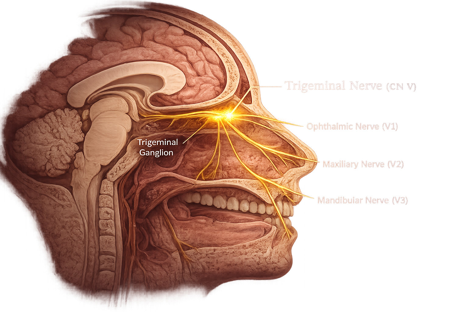
The trigeminal root emerges from the pons of the brainstem. It consists of a large sensory root and a smaller motor root. These roots transmit sensory information from the face and motor signals to the muscles of chewing.
The trigeminal ganglion, also called the semilunar or Gasserian ganglion, contains the cell bodies of sensory neurons. It serves as the main sensory relay station before the nerve divides into its three major branches.
The ophthalmic nerve (V1) is the first and purely sensory branch of the trigeminal nerve. It carries sensations from the forehead, scalp, upper eyelid, cornea, and the upper part of the nose to the brain.
The maxillary nerve (V2) is the second and purely sensory branch. It supplies sensation to the cheeks, lower eyelids, upper lip, upper teeth, nasal cavity, and the roof of the mouth.
The mandibular nerve (V3) is the third and only mixed branch of the trigeminal nerve. It provides sensation to the lower jaw, lower teeth, tongue, and chin, while also controlling the muscles responsible for chewing.
The motor fibers of the mandibular nerve supply the masseter, temporalis, medial pterygoid, and lateral pterygoid muscles. These muscles work together to open, close, and move the jaw during chewing and speaking.
The trigeminal nerve (CN V) is the largest cranial nerve and a mixed nerve with both sensory and motor functions. It provides sensation to most of the face and controls the muscles used for chewing, making it essential for everyday activities such as eating, speaking, and feeling facial sensations.
The abducens nerve (CN VI) is the sixth cranial nerve and is a motor nerve. It controls the lateral rectus muscle of the eye, which moves the eye outward (away from the nose). This nerve is essential for coordinated eye movements and proper side-to-side vision.
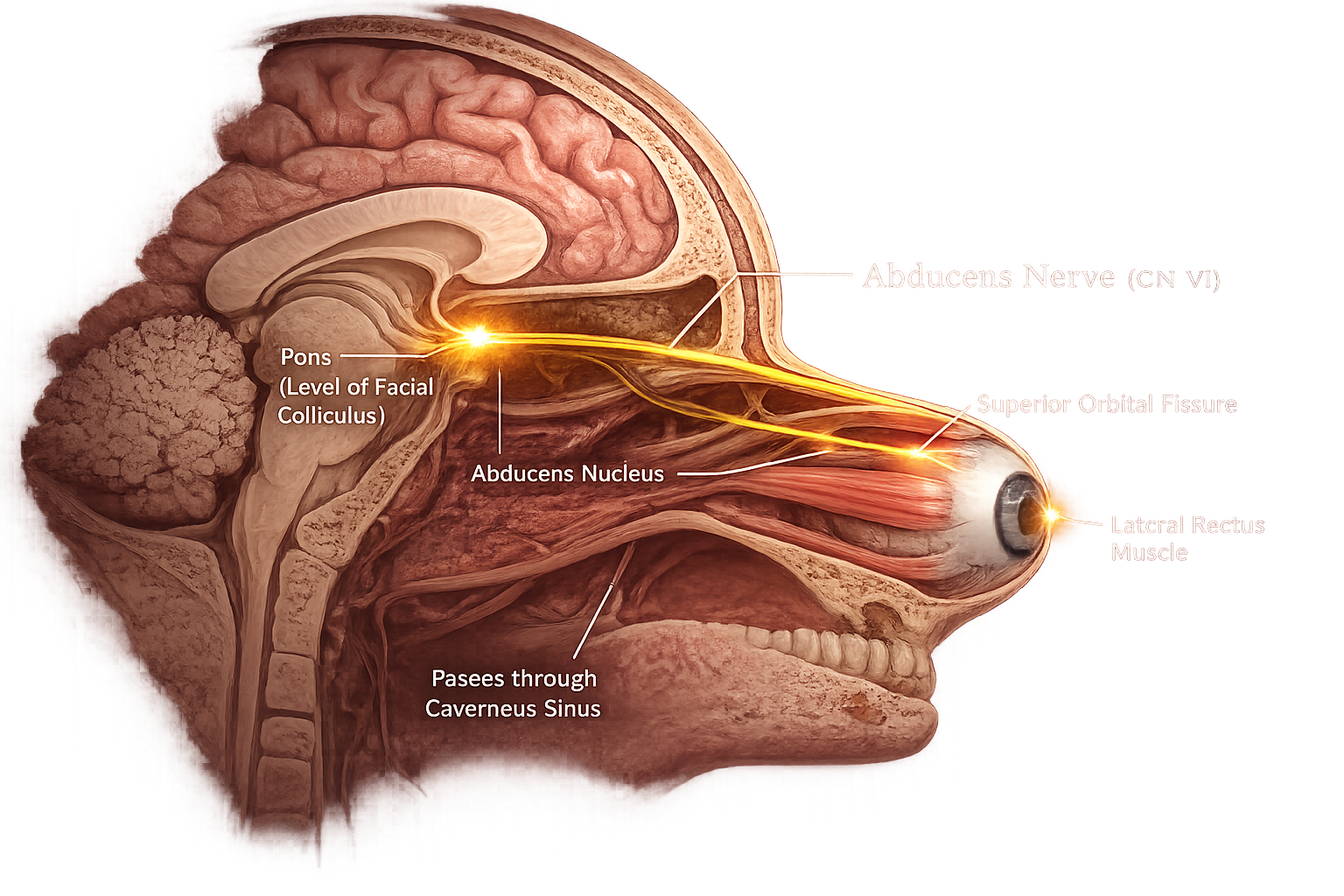
The abducens nucleus is located in the pons of the brainstem. It contains motor neurons that give rise to the abducens nerve and initiate the nerve impulses responsible for controlling the lateral rectus muscle of the eye.
The abducens nerve (CN VI) is a purely motor cranial nerve. It emerges from the junction between the pons and medulla oblongata before travelling toward the orbit to supply the lateral rectus muscle.
After leaving the brainstem, the abducens nerve passes through the cavernous sinus, a large venous space located on either side of the pituitary gland. It then continues toward the eye through the superior orbital fissure.
The superior orbital fissure is an opening in the skull through which the abducens nerve enters the orbit. It allows the nerve to reach the lateral rectus muscle of the eye.
The lateral rectus muscle is the only muscle supplied by the abducens nerve. It pulls the eyeball outward (laterally), allowing the eye to look away from the nose.
By controlling the lateral rectus muscle, the abducens nerve enables lateral eye movement (abduction). This movement is essential for coordinated vision, tracking moving objects, and maintaining binocular vision.
The abducens nerve (CN VI) is a motor nerve that controls the lateral rectus muscle of the eye. It enables the eye to move outward, helping maintain proper eye coordination, horizontal vision, and clear visual focus.
The facial nerve (CN VII) is the seventh cranial nerve and is a mixed nerve, containing both sensory and motor fibers. It controls the muscles responsible for facial expressions, provides the sense of taste from the front two-thirds of the tongue, and helps regulate the secretion of tears and saliva.
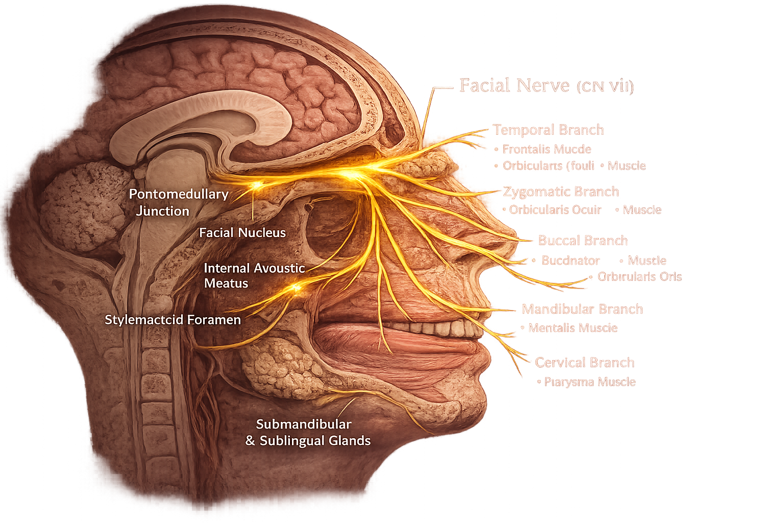
The facial nucleus is located in the pons of the brainstem. It contains motor neurons that give rise to the facial nerve and control the muscles responsible for facial expressions, such as smiling, frowning, and blinking.
The facial nerve (CN VII) is a mixed cranial nerve, carrying motor, sensory, and parasympathetic fibers. It emerges from the pontomedullary junction and travels through the temporal bone before branching across the face.
The internal acoustic meatus is a canal within the temporal bone through which the facial nerve enters the skull. Here, it travels alongside the vestibulocochlear nerve (CN VIII) before continuing through the facial canal.
The stylomastoid foramen is the opening through which the facial nerve exits the skull. After leaving this opening, it divides into several terminal branches that supply the muscles of facial expression.
The facial nerve divides into five main terminal branches: Temporal, Zygomatic, Buccal, Marginal Mandibular, and Cervical. These branches control the muscles of the forehead, eyelids, cheeks, lips, and neck, allowing a wide range of facial expressions.
The facial nerve carries parasympathetic fibers to the lacrimal gland and the submandibular and sublingual salivary glands. It also carries taste sensations from the anterior two-thirds of the tongue.
The facial nerve (CN VII) is a mixed cranial nerve that plays an essential role in facial expressions, taste, and the secretion of tears and saliva. It enables us to express emotions, enjoy the taste of food, and perform everyday activities such as speaking, blinking, and eating.
The vestibulocochlear nerve (CN VIII) is the eighth cranial nerve and is a sensory nerve. It is responsible for the senses of hearing and balance, carrying sound and equilibrium information from the inner ear to the brain.
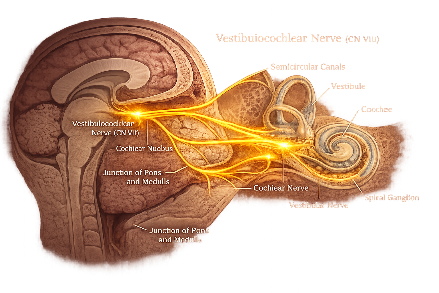
The vestibulocochlear nuclei are located at the junction of the pons and medulla oblongata. They receive sensory information from the inner ear and process signals related to hearing and balance before transmitting them to higher brain centers.
The vestibulocochlear nerve (CN VIII) is a purely sensory cranial nerve. It emerges from the brainstem and travels through the internal acoustic meatus to connect the brain with the inner ear, carrying impulses for hearing and equilibrium.
The cochlear nerve is the hearing division of CN VIII. It carries sound impulses from the cochlea to the brain, allowing the perception of speech, music, and environmental sounds.
The vestibular nerve is the balance division of CN VIII. It transmits information from the vestibule and semicircular canals to help maintain posture, balance, and coordinated head and eye movements.
The cochlea is the spiral-shaped organ of hearing in the inner ear. It contains specialized hair cells that convert sound vibrations into electrical nerve impulses, which are carried by the cochlear nerve.
The vestibular apparatus consists of the vestibule and semicircular canals. These structures detect head position, rotation, and movement, enabling the brain to maintain balance, posture, and stable vision during motion.
The vestibulocochlear nerve (CN VIII) is a sensory nerve that is essential for hearing and balance. It carries sound and equilibrium information from the inner ear to the brain, helping us hear clearly, maintain body balance, and coordinate our movements.
The glossopharyngeal nerve (CN IX) is the ninth cranial nerve and is a mixed nerve, containing both sensory and motor fibers. It plays an important role in taste, swallowing, saliva production, and helps regulate certain automatic body functions, such as blood pressure.
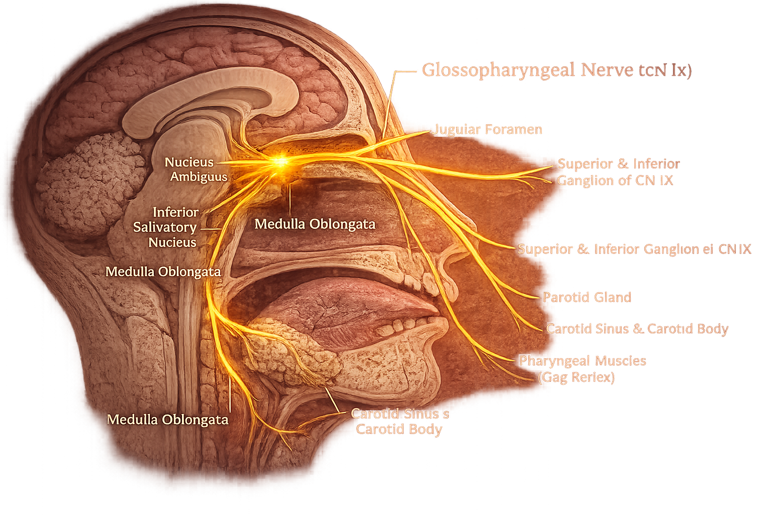
The glossopharyngeal nuclei are located in the medulla oblongata. These nuclei give rise to the glossopharyngeal nerve and contain sensory, motor, and parasympathetic neurons responsible for taste, swallowing, salivation, and reflexes.
The glossopharyngeal nerve (CN IX) is a mixed cranial nerve that carries sensory, motor, and parasympathetic fibers. It emerges from the medulla oblongata and exits the skull through the jugular foramen.
The jugular foramen is the opening in the base of the skull through which the glossopharyngeal nerve leaves the cranial cavity. It also serves as the exit point for the vagus nerve (CN X) and accessory nerve (CN XI).
The glossopharyngeal nerve provides taste sensation and general sensory information from the posterior one-third of the tongue, helping detect taste, touch, pain, and temperature.
The glossopharyngeal nerve carries parasympathetic fibers to the parotid salivary gland, stimulating the production and secretion of saliva during eating and digestion.
The stylopharyngeus muscle is the only skeletal muscle supplied by the glossopharyngeal nerve. It elevates the pharynx during swallowing and contributes to normal speech and swallowing movements.
The glossopharyngeal nerve (CN IX) is a mixed cranial nerve that plays an important role in taste, swallowing, and saliva production. It also helps regulate blood pressure and carries sensory information from the throat, making it essential for both voluntary and automatic body functions.
The vagus nerve (CN X) is the tenth cranial nerve and is a mixed nerve, containing both sensory and motor fibers. It is the longest cranial nerve and extends from the brainstem to the neck, chest, and abdomen. It plays a major role in regulating many automatic body functions, including heart rate, breathing, and digestion.
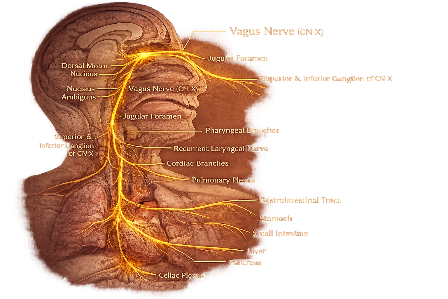
The vagus nuclei are located in the medulla oblongata. These nuclei contain motor, sensory, and parasympathetic neurons that control swallowing, speech, heart rate, breathing, and digestive functions.
The vagus nerve (CN X) is a mixed cranial nerve and the longest cranial nerve. It emerges from the medulla oblongata and travels through the neck into the thorax and abdomen, supplying numerous organs.
The jugular foramen is the opening through which the vagus nerve exits the skull. Shortly after passing through this foramen, it forms the superior and inferior sensory ganglia before descending into the neck.
The pharyngeal and recurrent laryngeal branches supply the muscles of the pharynx and larynx. They play essential roles in swallowing, speaking, and protecting the airway.
The vagus nerve gives off cardiac and pulmonary branches that regulate the heart and lungs. These parasympathetic fibers help slow the heart rate, regulate breathing, and maintain normal cardiovascular function.
The vagus nerve continues into the abdomen, supplying the stomach, liver, pancreas, small intestine, and other digestive organs. These branches stimulate digestion, promote gut movement, and regulate the secretion of digestive enzymes.
The vagus nerve (CN X) is the longest cranial nerve and a mixed nerve that controls many vital automatic body functions. It helps regulate heart rate, breathing, digestion, and swallowing, making it one of the most important nerves for maintaining the body's normal functions.
The accessory nerve (CN XI), also known as the spinal accessory nerve, is the eleventh cranial nerve and is a motor nerve. It controls the muscles responsible for head, neck, and shoulder movements, allowing actions such as turning the head and shrugging the shoulders.
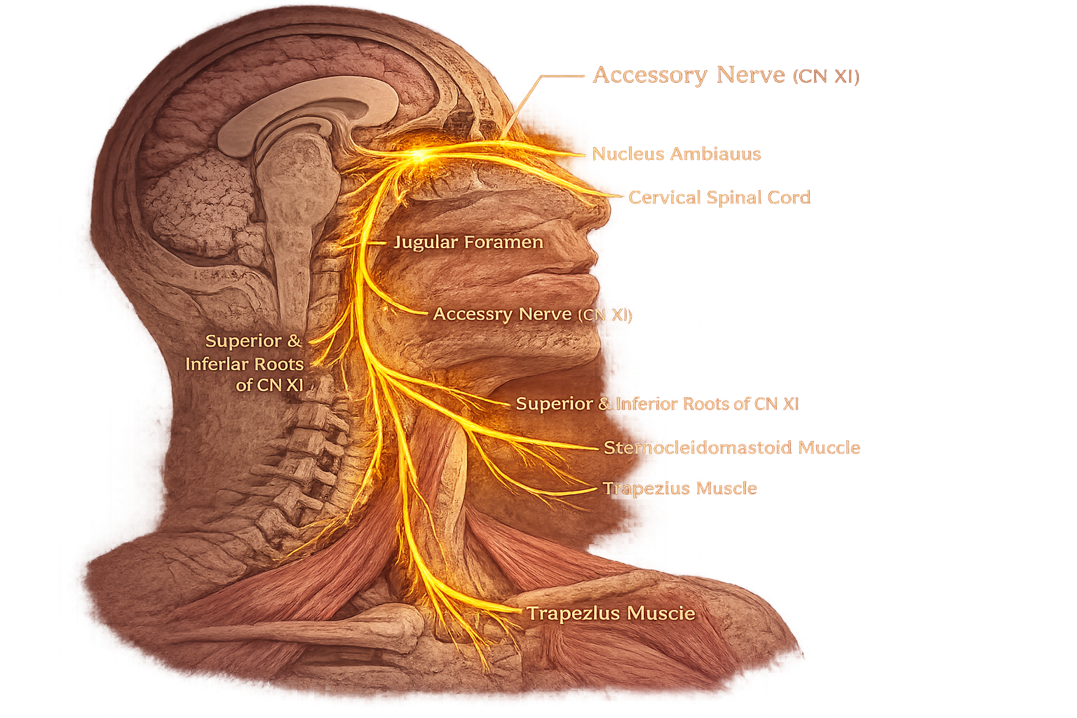
The accessory nucleus is located in the upper cervical spinal cord (C1–C5). It contains motor neurons that give rise to the spinal part of the accessory nerve, which controls important neck and shoulder muscles.
The accessory nerve (CN XI) is a purely motor cranial nerve. It is formed mainly by fibers arising from the cervical spinal cord, which enter the skull through the foramen magnum before exiting through the jugular foramen.
The jugular foramen is the opening at the base of the skull through which the accessory nerve exits the cranial cavity. It leaves alongside the glossopharyngeal (CN IX) and vagus (CN X) nerves before travelling into the neck.
The sternocleidomastoid muscle is one of the two muscles supplied by the accessory nerve. It allows the head to rotate from side to side and helps bend the neck forward during movement.
The trapezius muscle is the second muscle supplied by the accessory nerve. It elevates, retracts, and stabilizes the shoulders, making movements such as shrugging and lifting the arms possible.
Working together, the sternocleidomastoid and trapezius muscles enable smooth head rotation, neck movement, and shoulder elevation, which are essential for many everyday activities.
The accessory nerve (CN XI) is a motor nerve that plays an important role in controlling the movements of the head, neck, and shoulders. It enables actions such as turning the head and shrugging the shoulders, helping maintain proper posture and coordinated upper body movement.
The hypoglossal nerve (CN XII) is the twelfth cranial nerve and is a motor nerve. It controls the muscles of the tongue, allowing movements that are essential for speaking, chewing, and swallowing.
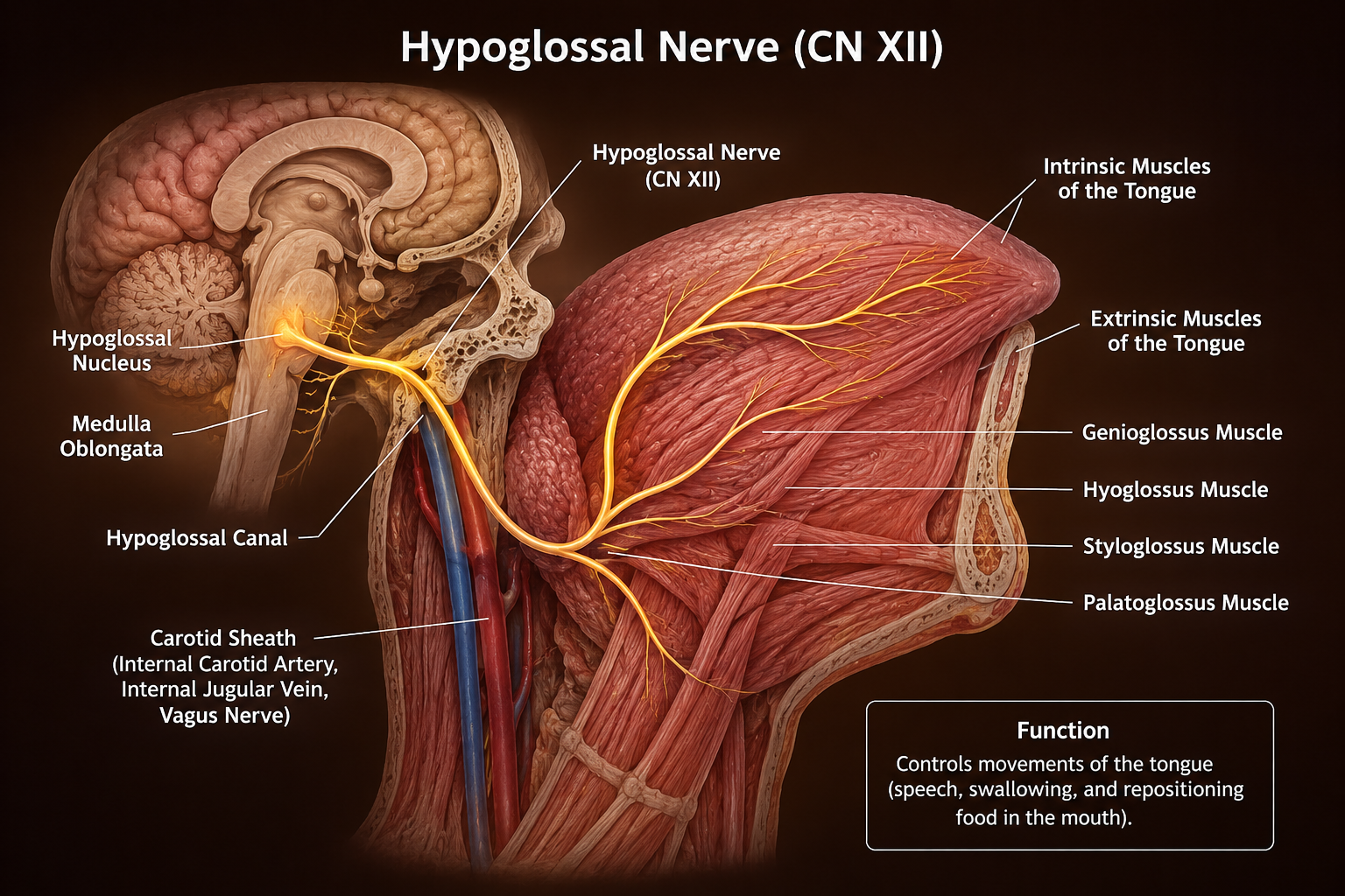
The hypoglossal nucleus is located in the medulla oblongata of the brainstem. It contains motor neurons that give rise to the hypoglossal nerve and control nearly all voluntary movements of the tongue.
The hypoglossal nerve (CN XII) is a purely motor cranial nerve. It emerges from the medulla oblongata and exits the skull through the hypoglossal canal before travelling to the tongue.
The hypoglossal canal is an opening in the occipital bone through which the hypoglossal nerve leaves the cranial cavity. It serves as the main passage connecting the brainstem to the tongue.
The intrinsic muscles are located entirely within the tongue. They change the shape of the tongue, allowing movements such as curling, flattening, shortening, and narrowing during speech and swallowing.
The hypoglossal nerve supplies the genioglossus, hyoglossus, and styloglossus muscles. These extrinsic muscles move the tongue in different directions, enabling protrusion, retraction, elevation, and depression.
By controlling the muscles of the tongue, the hypoglossal nerve allows precise movements required for speaking, chewing, swallowing, and moving food inside the mouth. It plays a vital role in clear speech and normal eating.
The hypoglossal nerve (CN XII) is a motor nerve that controls the movements of the tongue. It plays an essential role in speaking, chewing, and swallowing, ensuring proper tongue movement for effective communication and eating.